Structural and biophysical studies on two promoter recognition domains of the extra-cytoplasmic function sigma factor sigma(C) from Mycobacterium tuberculosis.
Thakur, K.G., Joshi, A.M., Gopal, B.(2007) J Biological Chem 282: 4711-4718
- PubMed: 17145760 Search on PubMedSearch on PubMed Central
- DOI: https://doi.org/10.1074/jbc.M606283200
- Primary Citation Related Structures:
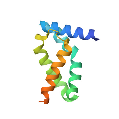
2O7G, 2O8X - PubMed Abstract:
sigma factors are transcriptional regulatory proteins that bind to the RNA polymerase and dictate gene expression. The extracytoplasmic function (ECF) sigma factors govern the environment dependent regulation of transcription. ECF sigma factors have two domains sigma(2) and sigma(4) that recognize the -10 and -35 promoter elements. However, unlike the primary sigma factor sigma(A), the ECF sigma factors lack sigma(3), a region that helps in the recognition of the extended -10 element and sigma(1.1), a domain involved in the autoinhibition of sigma(A) in the absence of core RNA polymerase. Mycobacterium tuberculosis sigma(C) is an ECF sigma factor that is essential for the pathogenesis and virulence of M. tuberculosis in the mouse and guinea pig models of infection. However, unlike other ECF sigma factors, sigma(C) does not appear to have a regulatory anti-sigma factor located in the same operon. We also note that M. tuberculosis sigma(C) differs from the canonical ECF sigma factors as it has an N-terminal domain comprising of 126 amino acids that precedes the sigma(C)(2) and sigma(C)(4) domains. In an effort to understand the regulatory mechanism of this protein, the crystal structures of the sigma(C)(2) and sigma(C)(4) domains of sigma(C) were determined. These promoter recognition domains are structurally similar to the corresponding domains of sigma(A) despite the low sequence similarity. Fluorescence experiments using the intrinsic tryptophan residues of sigma(C)(2) as well as surface plasmon resonance measurements reveal that the sigma(C)(2) and sigma(C)(4) domains interact with each other. Mutational analysis suggests that the Pribnow box-binding region of sigma(C)(2) is involved in this interdomain interaction. Interaction between the promoter recognition domains in M. tuberculosis sigma(C) are thus likely to regulate the activity of this protein even in the absence of an anti-sigma factor.
- Molecular Biophysics Unit, Indian Institute of Science, Bangalore 560 012, India.
Organizational Affiliation: