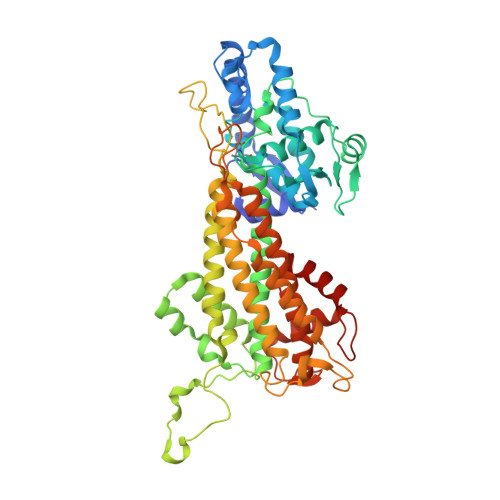
2O7B

Tyrosine ammonia-lyase from Rhodobacter sphaeroides, complexed with coumarate


Experimental Data Snapshot

  • Method: X-RAY DIFFRACTION
  • Resolution: 1.60 Å
  • R-Value Free: 0.208 
  • R-Value Work: 0.192 

wwPDB Validation   3D Report Full Report


Ligand Structure Quality Assessment 


This is version 2.0 of the entry. See complete history


Literature

Structural determinants and modulation of substrate specificity in phenylalanine-tyrosine ammonia-lyases.

Louie, G.V.Bowman, M.E.Moffitt, M.C.Baiga, T.J.Moore, B.S.Noel, J.P.

(2006) Chem Biol 13: 1327-1338

  • DOI: https://doi.org/10.1016/j.chembiol.2006.11.011
  • Primary Citation of Related Structures:  
    2O6Y, 2O78, 2O7B, 2O7D, 2O7E, 2O7F

  • PubMed Abstract: 

    Aromatic amino acid ammonia-lyases catalyze the deamination of L-His, L-Phe, and L-Tyr, yielding ammonia plus aryl acids bearing an alpha,beta-unsaturated propenoic acid. We report crystallographic analyses of unliganded Rhodobacter sphaeroides tyrosine ammonia-lyase (RsTAL) and RsTAL bound to p-coumarate and caffeate. His 89 of RsTAL forms a hydrogen bond with the p-hydroxyl moieties of coumarate and caffeate. His 89 is conserved in TALs but replaced in phenylalanine ammonia-lyases (PALs) and histidine ammonia-lyases (HALs). Substitution of His 89 by Phe, a characteristic residue of PALs, yields a mutant with a switch in kinetic preference from L-Tyr to L-Phe. Structures of the H89F mutant in complex with the PAL product, cinnamate, or the PAL-specific inhibitor, 2-aminoindan-2-phosphonate (AIP), support the role of position 89 as a specificity determinant in the family of aromatic amino acid ammonia-lyases and aminomutases responsible for beta-amino acid biosynthesis.


  • Organizational Affiliation

    Howard Hughes Medical Institute, Jack H. Skirball Center for Chemical Biology and Proteomics, The Salk Institute for Biological Studies, La Jolla, California 92037, USA.


Macromolecules
Find similar proteins by:  (by identity cutoff)  |  3D Structure
Entity ID: 1
MoleculeChains Sequence LengthOrganismDetailsImage
Putative histidine ammonia-lyase
A, B, C, D, E
A, B, C, D, E, F, G, H
521Cereibacter sphaeroidesMutation(s): 0 
Gene Names: hutH
EC: 4.3.1
UniProt
Find proteins for Q3IWB0 (Cereibacter sphaeroides (strain ATCC 17023 / DSM 158 / JCM 6121 / CCUG 31486 / LMG 2827 / NBRC 12203 / NCIMB 8253 / ATH 2.4.1.))
Explore Q3IWB0 
Go to UniProtKB:  Q3IWB0
Entity Groups  
Sequence Clusters30% Identity50% Identity70% Identity90% Identity95% Identity100% Identity
UniProt GroupQ3IWB0
Sequence Annotations
Expand
  • Reference Sequence
Experimental Data & Validation

Experimental Data

  • Method: X-RAY DIFFRACTION
  • Resolution: 1.60 Å
  • R-Value Free: 0.208 
  • R-Value Work: 0.192 
  • Space Group: P 1 21 1
Unit Cell:
Length ( Å )Angle ( ˚ )
a = 87.411α = 90
b = 154.823β = 94.11
c = 164.061γ = 90
Software Package:
Software NamePurpose
Blu-Icedata collection
CNSrefinement
XDSdata reduction
XSCALEdata scaling

Structure Validation

View Full Validation Report


Ligand Structure Quality Assessment 


Entry History 

Deposition Data

Revision History  (Full details and data files)

  • Version 1.0: 2007-01-16
    Type: Initial release
  • Version 1.1: 2008-05-01
    Changes: Version format compliance
  • Version 1.2: 2011-07-13
    Changes: Version format compliance
  • Version 1.3: 2023-08-30
    Changes: Data collection, Database references, Derived calculations, Refinement description
  • Version 2.0: 2023-11-15
    Changes: Atomic model, Data collection, Derived calculations