2O4V

An arginine ladder in OprP mediates phosphate specific transfer across the outer membrane


Experimental Data Snapshot

  • Method: X-RAY DIFFRACTION
  • Resolution: 1.94 Å
  • R-Value Free: 0.194 
  • R-Value Work: 0.174 
  • R-Value Observed: 0.175 

wwPDB Validation   3D Report Full Report


Ligand Structure Quality Assessment 


This is version 1.3 of the entry. See complete history


Literature

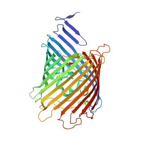
An arginine ladder in OprP mediates phosphate-specific transfer across the outer membrane.

Moraes, T.F.Bains, M.Hancock, R.E.Strynadka, N.C.

(2007) Nat Struct Mol Biol 14: 85-87

  • DOI: https://doi.org/10.1038/nsmb1189
  • Primary Citation of Related Structures:  
    2O4V

  • PubMed Abstract: 

    The outer membrane protein OprP mediates the transport of essential phosphate anions into the pathogenic bacterium Pseudomonas aeruginosa. Here we report the crystallographic structure of trimeric OprP at 1.9-A resolution, revealing an unprecedented 9-residue arginine 'ladder' that spans from the extracellular surface down through a constriction zone where phosphate is coordinated. Lysine residues coat the inner periplasmic surface, creating an 'electropositive sink' that pulls the phosphates through the eyelet and into the cell.


  • Organizational Affiliation

    Department of Biochemistry and Molecular Biology and the Center for Blood Research, University of British Columbia, 2350 Health Sciences Mall, Vancouver, British Columbia V6T 1Z3, Canada.


Macromolecules
Find similar proteins by:  (by identity cutoff)  |  3D Structure
Entity ID: 1
MoleculeChains Sequence LengthOrganismDetailsImage
Porin P
A, B, C
411Pseudomonas aeruginosa PAO1Mutation(s): 0 
Membrane Entity: Yes 
UniProt
Find proteins for P05695 (Pseudomonas aeruginosa (strain ATCC 15692 / DSM 22644 / CIP 104116 / JCM 14847 / LMG 12228 / 1C / PRS 101 / PAO1))
Explore P05695 
Go to UniProtKB:  P05695
Entity Groups  
Sequence Clusters30% Identity50% Identity70% Identity90% Identity95% Identity100% Identity
UniProt GroupP05695
Sequence Annotations
Expand
  • Reference Sequence
Small Molecules
Ligands 5 Unique
IDChains Name / Formula / InChI Key2D Diagram3D Interactions
C8E
Query on C8E

Download Ideal Coordinates CCD File 
G [auth A],
H [auth A],
I [auth A],
O [auth C]
(HYDROXYETHYLOXY)TRI(ETHYLOXY)OCTANE
C16 H34 O5
FEOZZFHAVXYAMB-UHFFFAOYSA-N
PO4
Query on PO4

Download Ideal Coordinates CCD File 
E [auth A],
L [auth B],
N [auth C]
PHOSPHATE ION
O4 P
NBIIXXVUZAFLBC-UHFFFAOYSA-K
GOL
Query on GOL

Download Ideal Coordinates CCD File 
J [auth A],
P [auth C]
GLYCEROL
C3 H8 O3
PEDCQBHIVMGVHV-UHFFFAOYSA-N
CA
Query on CA

Download Ideal Coordinates CCD File 
D [auth A],
K [auth B],
M [auth C]
CALCIUM ION
Ca
BHPQYMZQTOCNFJ-UHFFFAOYSA-N
CL
Query on CL

Download Ideal Coordinates CCD File 
F [auth A]CHLORIDE ION
Cl
VEXZGXHMUGYJMC-UHFFFAOYSA-M
Experimental Data & Validation

Experimental Data

  • Method: X-RAY DIFFRACTION
  • Resolution: 1.94 Å
  • R-Value Free: 0.194 
  • R-Value Work: 0.174 
  • R-Value Observed: 0.175 
  • Space Group: C 1 2 1
Unit Cell:
Length ( Å )Angle ( ˚ )
a = 255.13α = 90
b = 142.769β = 99.49
c = 62.025γ = 90
Software Package:
Software NamePurpose
REFMACrefinement
ADSCdata collection
MOSFLMdata reduction
CCP4data scaling
SHELXSphasing

Structure Validation

View Full Validation Report


Ligand Structure Quality Assessment 


Entry History 

Deposition Data

Revision History  (Full details and data files)

  • Version 1.0: 2006-12-26
    Type: Initial release
  • Version 1.1: 2008-05-01
    Changes: Version format compliance
  • Version 1.2: 2011-07-13
    Changes: Non-polymer description, Version format compliance
  • Version 1.3: 2023-12-27
    Changes: Data collection, Database references, Derived calculations