2O4J

Crystal Structure of Rat Vitamin D Receptor Ligand Binding Domain Complexed with VitIII 17-20Z and the NR2 Box of DRIP 205


Experimental Data Snapshot

  • Method: X-RAY DIFFRACTION
  • Resolution: 1.74 Å
  • R-Value Free: 0.246 
  • R-Value Work: 0.199 
  • R-Value Observed: 0.201 

wwPDB Validation   3D Report Full Report


Ligand Structure Quality Assessment 


This is version 1.4 of the entry. See complete history


Literature

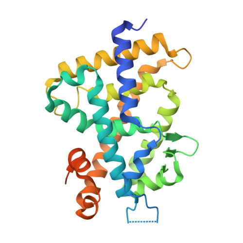
New analogs of 2-methylene-19-nor-(20S)-1,25-dihydroxyvitamin D(3) with conformationally restricted side chains: Evaluation of biological activity and structural determination of VDR-bound conformations.

Vanhooke, J.L.Tadi, B.P.Benning, M.M.Plum, L.A.Deluca, H.F.

(2007) Arch Biochem Biophys 460: 161-165

  • DOI: https://doi.org/10.1016/j.abb.2006.11.029
  • Primary Citation of Related Structures:  
    2O4J, 2O4R

  • PubMed Abstract: 
  • We have successfully prepared E- and Z- isomers of 17-20 dehydro analogs of 2-methylene-19-nor-(20S)-1alpha,25-dihydroxyvitamin D3 (2MD). Both isomers bind to the recombinant rat vitamin D receptor (VDR) with high affinity. The Z-isomer (Vit-III 17-20Z) displays activity in vivo and in vitro that is similar to 2MD ...

    We have successfully prepared E- and Z- isomers of 17-20 dehydro analogs of 2-methylene-19-nor-(20S)-1alpha,25-dihydroxyvitamin D3 (2MD). Both isomers bind to the recombinant rat vitamin D receptor (VDR) with high affinity. The Z-isomer (Vit-III 17-20Z) displays activity in vivo and in vitro that is similar to 2MD. The in vitro activity of the E-isomer (Vit-III 17-20E) is comparable to the natural hormone, though in vivo this analog is significantly less calcemic. Crystal structures of the rat VDR ligand binding domain complexed with the analogs demonstrate that the Vit-III 17-20Z analog is oriented almost identically to 2MD, with only minor differences induced by the planar configuration around the C17-C20 double bond. The Vit-III 17-20E analog is oriented in a conformation distinct from both 2MD and the natural hormone. The structural comparisons suggest that the position of C21 in the ligand binding site may be an important determinant of biological activity.


    Related Citations: 
    • Molecular Structure of the Rat Vitamin D Receptor Ligand Binding Domain Complexed with 2-Carbon-Substituted Vitamin D3 Hormone Analogues and a LXXLL-Containing Coactivator Peptide
      Vanhooke, J.L., Benning, M.M., Bauer, C.B., Pike, J.W., DeLuca, H.F.
      (2004) Biochemistry 43: 4101

    Organizational Affiliation

    Department of Biochemistry, University of Wisconsin-Madison, 433 Babcock Drive, Madison, WI 53706, USA. vanhooke@med.unc.edu


Macromolecules
Find similar proteins by:  (by identity cutoff)  |  3D Structure
Entity ID: 1
MoleculeChainsSequence LengthOrganismDetailsImage
Vitamin D3 receptor292Rattus norvegicusMutation(s): 0 
Gene Names: VdrNr1i1
UniProt
Find proteins for P13053 (Rattus norvegicus)
Explore P13053 
Go to UniProtKB:  P13053
Entity Groups  
Sequence Clusters30% Identity50% Identity70% Identity90% Identity95% Identity100% Identity
UniProt GroupP13053
Protein Feature View
Expand
  • Reference Sequence

Find similar proteins by:  Sequence   |   3D Structure  

Entity ID: 2
MoleculeChainsSequence LengthOrganismDetailsImage
Peroxisome proliferator-activated receptor-binding proteinB [auth C]13N/AMutation(s): 0 
UniProt & NIH Common Fund Data Resources
Find proteins for Q15648 (Homo sapiens)
Explore Q15648 
Go to UniProtKB:  Q15648
PHAROS:  Q15648
Entity Groups  
Sequence Clusters30% Identity50% Identity70% Identity90% Identity95% Identity100% Identity
UniProt GroupQ15648
Protein Feature View
Expand
  • Reference Sequence
Small Molecules
Ligands 1 Unique
IDChainsName / Formula / InChI Key2D Diagram3D Interactions
VD4
Query on VD4

Download Ideal Coordinates CCD File 
C [auth A](1R,3R,7E,17Z)-17-(5-hydroxy-1,5-dimethylhexylidene)-2-methylene-9,10-secoestra-5,7-diene-1,3-diol
C27 H42 O3
PCPYFQNISYYIPU-DUKKOGHCSA-N
 Ligand Interaction
Binding Affinity Annotations 
IDSourceBinding Affinity
VD4 Binding MOAD:  2O4J Ki: 0.08 (nM) from 1 assay(s)
PDBBind:  2O4J Ki: 0.08 (nM) from 1 assay(s)
Experimental Data & Validation

Experimental Data

  • Method: X-RAY DIFFRACTION
  • Resolution: 1.74 Å
  • R-Value Free: 0.246 
  • R-Value Work: 0.199 
  • R-Value Observed: 0.201 
  • Space Group: C 1 2 1
Unit Cell:
Length ( Å )Angle ( ˚ )
a = 155.5α = 90
b = 42.4β = 96.5
c = 41.9γ = 90
Software Package:
Software NamePurpose
REFMACrefinement
PDB_EXTRACTdata extraction
SAINTdata reduction
LSCALEdata scaling

Structure Validation

View Full Validation Report


Ligand Structure Quality Assessment 


Entry History 

Deposition Data

Revision History  (Full details and data files)

  • Version 1.0: 2007-01-30
    Type: Initial release
  • Version 1.1: 2008-05-01
    Changes: Version format compliance
  • Version 1.2: 2011-07-13
    Changes: Version format compliance
  • Version 1.3: 2017-10-18
    Changes: Refinement description
  • Version 1.4: 2023-08-30
    Changes: Data collection, Database references, Derived calculations, Refinement description