2O3T

Structural Basis for Formation and Hydrolysis of Calcium Messenger Cyclic ADP-ribose by Human CD38


Experimental Data Snapshot

  • Method: X-RAY DIFFRACTION
  • Resolution: 1.68 Å
  • R-Value Free: 0.208 
  • R-Value Work: 0.172 
  • R-Value Observed: 0.174 

wwPDB Validation   3D Report Full Report


This is version 1.4 of the entry. See complete history


Literature

Structural basis for formation and hydrolysis of the calcium messenger cyclic ADP-ribose by human CD38

Liu, Q.Kriksunov, I.A.Graeff, R.Lee, H.C.Hao, Q.

(2007) J Biol Chem 282: 5853-5861

  • DOI: https://doi.org/10.1074/jbc.M609093200
  • Primary Citation of Related Structures:  
    2O3Q, 2O3R, 2O3S, 2O3T, 2O3U

  • PubMed Abstract: 

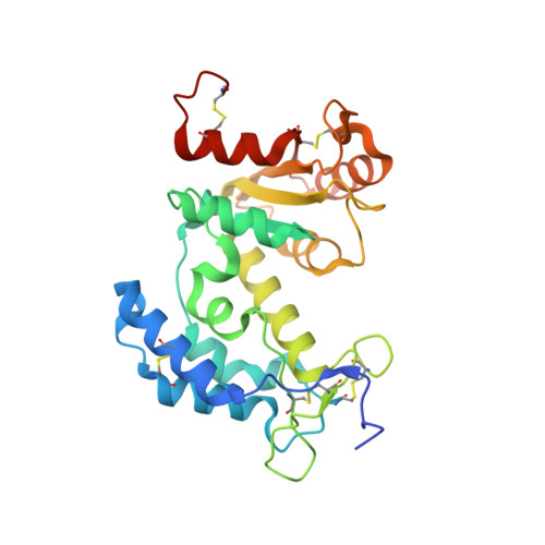
    Human CD38 is a multifunctional ectoenzyme responsible for catalyzing the conversions from nicotinamide adenine dinucleotide (NAD) to cyclic ADP-ribose (cADPR) and from cADPR to ADP-ribose (ADPR). Both cADPR and ADPR are calcium messengers that can mobilize intracellular stores and activate influx as well. In this study, we determined three crystal structures of the human CD38 enzymatic domain complexed with cADPR at 1.5-A resolution, with its analog, cyclic GDP-ribose (cGDPR) (1.68 A) and with NGD (2.1 A) a substrate analog of NAD. The results indicate that the binding of cADPR or cGDPR to the active site induces structural rearrangements in the dipeptide Glu(146)-Asp(147) by as much as 2.7 A) providing the first direct evidence of a conformational change at the active site during catalysis. In addition, Glu(226) is shown to be critical not only in catalysis but also in positioning of cADPR at the catalytic site through strong hydrogen bonding interactions. Structural details obtained from these complexes provide a step-by-step description of the catalytic processes in the synthesis and hydrolysis of cADPR.


  • Organizational Affiliation

    MacCHESS, Cornell High Energy Synchrotron Source, Cornell University, Ithaca, New York 14853, USA.


Macromolecules
Find similar proteins by:  (by identity cutoff)  |  3D Structure
Entity ID: 1
MoleculeChains Sequence LengthOrganismDetailsImage
ADP-ribosyl cyclase 1
A, B
262Homo sapiensMutation(s): 6 
Gene Names: CD38
EC: 3.2.2.5
UniProt & NIH Common Fund Data Resources
Find proteins for P28907 (Homo sapiens)
Explore P28907 
Go to UniProtKB:  P28907
PHAROS:  P28907
GTEx:  ENSG00000004468 
Entity Groups  
Sequence Clusters30% Identity50% Identity70% Identity90% Identity95% Identity100% Identity
UniProt GroupP28907
Sequence Annotations
Expand
  • Reference Sequence
Small Molecules
Ligands 1 Unique
IDChains Name / Formula / InChI Key2D Diagram3D Interactions
CGR
Query on CGR

Download Ideal Coordinates CCD File 
C [auth A]CYCLIC GUANOSINE DIPHOSPHATE-RIBOSE
C15 H23 N5 O14 P2
XBIQPWWXYHHVOT-AIKIOGRUSA-N
Experimental Data & Validation

Experimental Data

  • Method: X-RAY DIFFRACTION
  • Resolution: 1.68 Å
  • R-Value Free: 0.208 
  • R-Value Work: 0.172 
  • R-Value Observed: 0.174 
  • Space Group: P 1
Unit Cell:
Length ( Å )Angle ( ˚ )
a = 41.994α = 105.9
b = 53.289β = 91.76
c = 65.729γ = 95.16
Software Package:
Software NamePurpose
REFMACrefinement
ADSCdata collection
HKL-3000data reduction
HKL-3000data scaling
MOLREPphasing

Structure Validation

View Full Validation Report


Entry History 

Deposition Data

Revision History  (Full details and data files)

  • Version 1.0: 2006-12-19
    Type: Initial release
  • Version 1.1: 2008-05-01
    Changes: Version format compliance
  • Version 1.2: 2011-07-13
    Changes: Version format compliance
  • Version 1.3: 2021-10-20
    Changes: Database references, Derived calculations
  • Version 1.4: 2023-08-30
    Changes: Data collection, Refinement description