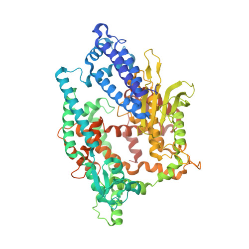
Swapping the substrate specificities of the neuropeptidases neurolysin and thimet oligopeptidase.
Lim, E.J., Sampath, S., Coll-Rodriguez, J., Schmidt, J., Ray, K., Rodgers, D.W.(2007) J Biological Chem 282: 9722-9732
- PubMed: 17251185
- DOI: https://doi.org/10.1074/jbc.M609897200
- Primary Citation of Related Structures:
2O36, 2O3E - PubMed Abstract:
Thimet oligopeptidase (EC 3.4.24.15) and neurolysin (EC 3.4.24.16) are closely related zinc-dependent metallopeptidases that metabolize small bioactive peptides. They cleave many substrates at the same sites, but they recognize different positions on others, including neurotensin, a 13-residue peptide involved in modulation of dopaminergic circuits, pain perception, and thermoregulation. On the basis of crystal structures and previous mapping studies, four sites (Glu-469/Arg-470, Met-490/Arg-491, His-495/Asn-496, and Arg-498/Thr-499; thimet oligopeptidase residues listed first) in their substrate-binding channels appear positioned to account for differences in specificity. Thimet oligopeptidase mutated so that neurolysin residues are at all four positions cleaves neurotensin at the neurolysin site, and the reverse mutations in neurolysin switch hydrolysis to the thimet oligopeptidase site. Using a series of constructs mutated at just three of the sites, it was determined that mutations at only two (Glu-469/Arg-470 and Arg-498/Thr-499) are required to swap specificity, a result that was confirmed by testing the two-mutant constructs. If only either one of the two sites is mutated in thimet oligopeptidase, then the enzyme cleaves almost equally at the two hydrolysis positions. Crystal structures of both two-mutant constructs show that the mutations do not perturb local structure, but side chain conformations at the Arg-498/Thr-499 position differ from those of the mimicked enzyme. A model for differential recognition of neurotensin based on differences in surface charge distribution in the substrate binding sites is proposed. The model is supported by the finding that reducing the positive charge on the peptide results in cleavage at both hydrolysis sites.
- Department of Molecular and Cellular Biochemistry and Center for Structural Biology, University of Kentucky, Lexington, Kentucky 40536.
Organizational Affiliation:


