2NVG

Soluble domain of Rieske Iron Sulfur protein.


Experimental Data Snapshot

  • Method: X-RAY DIFFRACTION
  • Resolution: 1.35 Å
  • R-Value Free: 0.159 
  • R-Value Work: 0.142 
  • R-Value Observed: 0.143 

wwPDB Validation   3D Report Full Report


This is version 1.4 of the entry. See complete history


Literature

Atomic resolution structures of rieske iron-sulfur protein: role of hydrogen bonds in tuning the redox potential of iron-sulfur clusters.

Kolling, D.J.Brunzelle, J.S.Lhee, S.Crofts, A.R.Nair, S.K.

(2007) Structure 15: 29-38

  • DOI: https://doi.org/10.1016/j.str.2006.11.012
  • Primary Citation of Related Structures:  
    2NUK, 2NUM, 2NVE, 2NVF, 2NVG, 2NWF

  • PubMed Abstract: 

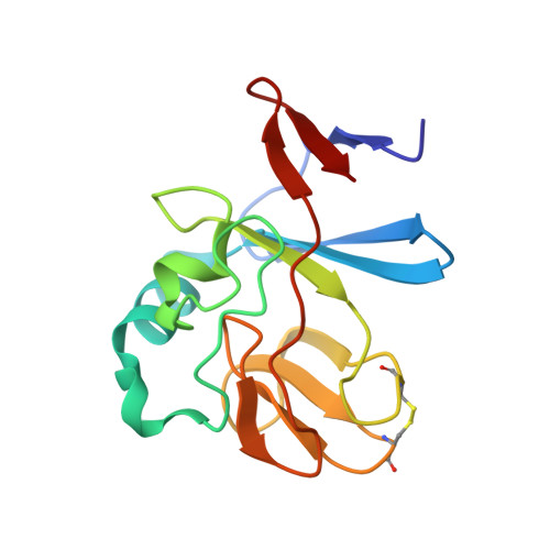
    The Rieske [2Fe-2S] iron-sulfur protein of cytochrome bc(1) functions as the initial electron acceptor in the rate-limiting step of the catalytic reaction. Prior studies have established roles for a number of conserved residues that hydrogen bond to ligands of the [2Fe-2S] cluster. We have constructed site-specific variants at two of these residues, measured their thermodynamic and functional properties, and determined atomic resolution X-ray crystal structures for the native protein at 1.2 A resolution and for five variants (Ser-154-->Ala, Ser-154-->Thr, Ser-154-->Cys, Tyr-156-->Phe, and Tyr-156-->Trp) to resolutions between 1.5 A and 1.1 A. These structures and complementary biophysical data provide a molecular framework for understanding the role hydrogen bonds to the cluster play in tuning thermodynamic properties, and hence the rate of this bioenergetic reaction. These studies provide a detailed structure-function dissection of the role of hydrogen bonds in tuning the redox potentials of [2Fe-2S] clusters.


  • Organizational Affiliation

    Center for Biophysics and Computational Biology, University of Illinois at Urbana-Champaign, 600 S. Mathews Avenue, Urbana, IL 61801, USA.


Macromolecules
Find similar proteins by:  (by identity cutoff)  |  3D Structure
Entity ID: 1
MoleculeChains Sequence LengthOrganismDetailsImage
Ubiquinol-cytochrome c reductase iron-sulfur subunit141Cereibacter sphaeroidesMutation(s): 1 
Gene Names: petAfbcF
EC: 1.10.2.2
UniProt
Find proteins for Q02762 (Cereibacter sphaeroides)
Explore Q02762 
Go to UniProtKB:  Q02762
Entity Groups  
Sequence Clusters30% Identity50% Identity70% Identity90% Identity95% Identity100% Identity
UniProt GroupQ02762
Sequence Annotations
Expand
  • Reference Sequence
Experimental Data & Validation

Experimental Data

  • Method: X-RAY DIFFRACTION
  • Resolution: 1.35 Å
  • R-Value Free: 0.159 
  • R-Value Work: 0.142 
  • R-Value Observed: 0.143 
  • Space Group: I 4
Unit Cell:
Length ( Å )Angle ( ˚ )
a = 69.935α = 90
b = 69.935β = 90
c = 54.582γ = 90
Software Package:
Software NamePurpose
REFMACrefinement
ADSCdata collection
HKL-2000data reduction
HKL-2000data scaling
MOLREPphasing

Structure Validation

View Full Validation Report


Entry History 

Deposition Data

Revision History  (Full details and data files)

  • Version 1.0: 2007-02-06
    Type: Initial release
  • Version 1.1: 2008-05-01
    Changes: Version format compliance
  • Version 1.2: 2011-07-13
    Changes: Non-polymer description, Version format compliance
  • Version 1.3: 2021-10-20
    Changes: Database references, Derived calculations
  • Version 1.4: 2023-08-30
    Changes: Data collection, Refinement description