2NRM

S-nitrosylated blackfin tuna myoglobin


Experimental Data Snapshot

  • Method: X-RAY DIFFRACTION
  • Resolution: 1.09 Å
  • R-Value Free: 0.157 
  • R-Value Work: 0.133 
  • R-Value Observed: 0.134 

wwPDB Validation   3D Report Full Report


Ligand Structure Quality Assessment 


This is version 1.4 of the entry. See complete history


Literature

S-nitrosylation-induced conformational change in blackfin tuna myoglobin.

Schreiter, E.R.Rodriguez, M.M.Weichsel, A.Montfort, W.R.Bonaventura, J.

(2007) J Biol Chem 282: 19773-19780

  • DOI: https://doi.org/10.1074/jbc.M701363200
  • Primary Citation of Related Structures:  
    2NRL, 2NRM, 2NX0

  • PubMed Abstract: 

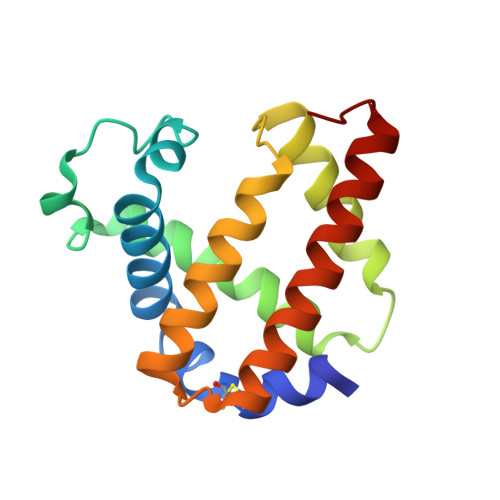
    S-nitrosylation is a post-translational protein modification that can alter the function of a variety of proteins. Despite the growing wealth of information that this modification may have important functional consequences, little is known about the structure of the moiety or its effect on protein tertiary structure. Here we report high-resolution x-ray crystal structures of S-nitrosylated and unmodified blackfin tuna myoglobin, which demonstrate that in vitro S-nitrosylation of this protein at the surface-exposed Cys-10 directly causes a reversible conformational change by "wedging" apart a helix and loop. Furthermore, we have demonstrated in solution and in a single crystal that reduction of the S-nitrosylated myoglobin with dithionite results in NO cleavage from the sulfur of Cys-10 and rebinding to the reduced heme iron, showing the reversibility of both the modification and the conformational changes. Finally, we report the 0.95-A structure of ferrous nitrosyl myoglobin, which provides an accurate structural view of the NO coordination geometry in the context of a globin heme pocket.


  • Organizational Affiliation

    Protein Research Center, Department of Chemistry, University of Puerto Rico, Mayagüez, Puerto Rico 00681. eric_sch@mit.edu


Macromolecules
Find similar proteins by:  (by identity cutoff)  |  3D Structure
Entity ID: 1
MoleculeChains Sequence LengthOrganismDetailsImage
Myoglobin147Thunnus atlanticusMutation(s): 0 
UniProt
Find proteins for P68189 (Thunnus thynnus)
Explore P68189 
Go to UniProtKB:  P68189
Entity Groups  
Sequence Clusters30% Identity50% Identity70% Identity90% Identity95% Identity100% Identity
UniProt GroupP68189
Sequence Annotations
Expand
  • Reference Sequence
Experimental Data & Validation

Experimental Data

  • Method: X-RAY DIFFRACTION
  • Resolution: 1.09 Å
  • R-Value Free: 0.157 
  • R-Value Work: 0.133 
  • R-Value Observed: 0.134 
  • Space Group: P 1 21 1
Unit Cell:
Length ( Å )Angle ( ˚ )
a = 33.943α = 90
b = 51.932β = 103.75
c = 38.899γ = 90
Software Package:
Software NamePurpose
REFMACrefinement
CrystalCleardata collection
CrystalCleardata reduction
CrystalCleardata scaling
PHASERphasing

Structure Validation

View Full Validation Report


Ligand Structure Quality Assessment 


Entry History 

Deposition Data

Revision History  (Full details and data files)

  • Version 1.0: 2007-05-08
    Type: Initial release
  • Version 1.1: 2007-10-23
    Changes: Version format compliance
  • Version 1.2: 2011-07-13
    Changes: Non-polymer description, Version format compliance
  • Version 1.3: 2018-04-04
    Changes: Advisory, Data collection
  • Version 1.4: 2023-08-30
    Changes: Advisory, Data collection, Database references, Derived calculations, Refinement description