2NPX

NADH BINDING SITE AND CATALYSIS OF NADH PEROXIDASE


Experimental Data Snapshot

  • Method: X-RAY DIFFRACTION
  • Resolution: 2.40 Å
  • R-Value Work: 0.159 
  • R-Value Observed: 0.159 

wwPDB Validation   3D Report Full Report


Ligand Structure Quality Assessment 


This is version 1.2 of the entry. See complete history


Literature

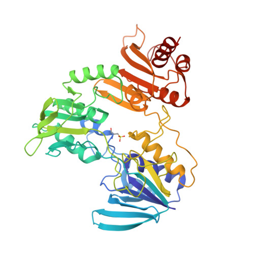
NADH binding site and catalysis of NADH peroxidase.

Stehle, T.Claiborne, A.Schulz, G.E.

(1993) Eur J Biochem 211: 221-226

  • DOI: https://doi.org/10.1111/j.1432-1033.1993.tb19889.x
  • Primary Citation of Related Structures:  
    2NPX

  • PubMed Abstract: 

    The structure of the complex between cofactor NADH and the enzyme NADH peroxidase from Streptococcus faecalis 10C1 (Enterococcus faecalis) has been determined by crystal soaking, X-ray data collection, model building of NADH and refinement at 0.24-nm resolution based on the known enzyme structure [Stehle, T., Ahmed, S. A., Claiborne, A. & Schulz, G. E. (1991) J. Mol. Biol. 221, 1325-1344]. Apart from NADH, the catalytic center of the enzyme contains FAD and a cysteine that shuttles between thiolate and sulfenic acid states. Unfortunately, this cysteine was irreversibly oxidized to a cysteine sulfonic acid in the established enzyme structure. Based on the geometry of the catalytic center, we discuss the stabilization of the oxidation-sensitive sulfenic acid and propose a reaction mechanism.


  • Organizational Affiliation

    Institut für Organische Chemie und Biochemie der Universität, Freiburg im Breisgau, Federal Republic of Germany.


Macromolecules
Find similar proteins by:  (by identity cutoff)  |  3D Structure
Entity ID: 1
MoleculeChains Sequence LengthOrganismDetailsImage
NADH PEROXIDASE447Enterococcus faecalisMutation(s): 0 
EC: 1.11.1.1
UniProt
Find proteins for P37062 (Enterococcus faecalis (strain ATCC 700802 / V583))
Explore P37062 
Go to UniProtKB:  P37062
Entity Groups  
Sequence Clusters30% Identity50% Identity70% Identity90% Identity95% Identity100% Identity
UniProt GroupP37062
Sequence Annotations
Expand
  • Reference Sequence
Small Molecules
Ligands 2 Unique
IDChains Name / Formula / InChI Key2D Diagram3D Interactions
FAD
Query on FAD

Download Ideal Coordinates CCD File 
B [auth A]FLAVIN-ADENINE DINUCLEOTIDE
C27 H33 N9 O15 P2
VWWQXMAJTJZDQX-UYBVJOGSSA-N
NAD
Query on NAD

Download Ideal Coordinates CCD File 
C [auth A]NICOTINAMIDE-ADENINE-DINUCLEOTIDE
C21 H27 N7 O14 P2
BAWFJGJZGIEFAR-NNYOXOHSSA-N
Modified Residues  1 Unique
IDChains TypeFormula2D DiagramParent
OCS
Query on OCS
A
L-PEPTIDE LINKINGC3 H7 N O5 SCYS
Experimental Data & Validation

Experimental Data

  • Method: X-RAY DIFFRACTION
  • Resolution: 2.40 Å
  • R-Value Work: 0.159 
  • R-Value Observed: 0.159 
  • Space Group: I 2 2 2
Unit Cell:
Length ( Å )Angle ( ˚ )
a = 77.2α = 90
b = 134.5β = 90
c = 145.9γ = 90
Software Package:
Software NamePurpose
X-PLORmodel building
X-PLORrefinement
X-PLORphasing

Structure Validation

View Full Validation Report


Ligand Structure Quality Assessment 


Entry History 

Deposition Data

Revision History  (Full details and data files)

  • Version 1.0: 1994-01-31
    Type: Initial release
  • Version 1.1: 2008-03-24
    Changes: Version format compliance
  • Version 1.2: 2011-07-13
    Changes: Derived calculations, Version format compliance