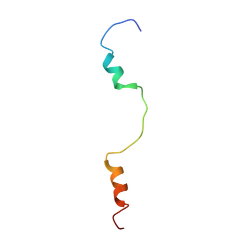
Solution Structure of Calmodulin Bound to the Binding Domain of the HIV-1 Matrix Protein.
Vlach, J., Samal, A.B., Saad, J.S.(2014) J Biological Chem 289: 8697-8705
- PubMed: 24500712
- DOI: https://doi.org/10.1074/jbc.M113.543694
- Primary Citation of Related Structures:
2MGU - PubMed Abstract:
Subcellular distribution of calmodulin (CaM) in human immunodeficiency virus type-1 (HIV-1)-infected cells is distinct from that observed in uninfected cells. CaM co-localizes and interacts with the HIV-1 Gag protein in the cytosol of infected cells. Although it has been shown that binding of Gag to CaM is mediated by the matrix (MA) domain, the structural details of this interaction are not known. We have recently shown that binding of CaM to MA induces a conformational change that triggers myristate exposure, and that the CaM-binding domain of MA is confined to a region spanning residues 8-43 (MA-(8-43)). Here, we present the NMR structure of CaM bound to MA-(8-43). Our data revealed that MA-(8-43), which contains a novel CaM-binding motif, binds to CaM in an antiparallel mode with the N-terminal helix (α1) anchored to the CaM C-terminal lobe, and the C-terminal helix (α2) of MA-(8-43) bound to the N-terminal lobe of CaM. The CaM protein preserves a semiextended conformation. Binding of MA-(8-43) to CaM is mediated by numerous hydrophobic interactions and stabilized by favorable electrostatic contacts. Our structural data are consistent with the findings that CaM induces unfolding of the MA protein to have access to helices α1 and α2. It is noteworthy that several MA residues involved in CaM binding have been previously implicated in membrane binding, envelope incorporation, and particle production. The present findings may ultimately help in identification of the functional role of CaM in HIV-1 replication.
- From the Department of Microbiology, University of Alabama at Birmingham, Birmingham, Alabama 35294.
Organizational Affiliation:


