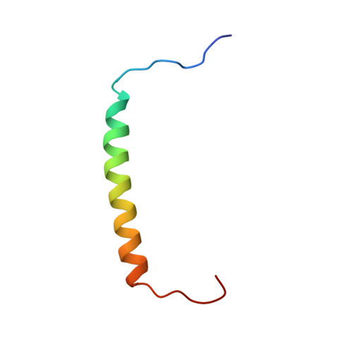
Alternative packing of EGFR transmembrane domain suggests that protein-lipid interactions underlie signal conduction across membrane.
Bocharov, E.V., Lesovoy, D.M., Pavlov, K.V., Pustovalova, Y.E., Bocharova, O.V., Arseniev, A.S.(2016) Biochim Biophys Acta 1858: 1254-1261
- PubMed: 26903218 Search on PubMed
- DOI: https://doi.org/10.1016/j.bbamem.2016.02.023
- Primary Citation Related Structures:
2M0B - PubMed Abstract:
The human epidermal growth factor receptor (EGFR) of HER/ErbB receptor tyrosine kinase family mediates a broad spectrum of cellular responses transducing biochemical signals via lateral dimerization in plasma membrane, while inactive receptors can exist in both monomeric and dimeric forms. Recently, the dimeric conformation of the helical single-span transmembrane domains of HER/ErbB employing the relatively polar N-terminal motifs in a fashion permitting proper kinase activation was experimentally determined. Here we describe the EGFR transmembrane domain dimerization via an alternative weakly polar C-terminal motif A(661)xxxG(665) presumably corresponding to the inactive receptor state. During association, the EGFR transmembrane helices undergo a structural adjustment with adaptation of inter-molecular polar and hydrophobic interactions depending upon the surrounding membrane properties that directly affect the transmembrane helix packing. This might imply that signal transduction through membrane and allosteric regulation are inclusively mediated by coupled protein-protein and protein-lipid interactions, elucidating paradoxically loose linkage between ligand binding and kinase activation.
- Department of Structural Biology, Shemyakin-Ovchinnikov Institute of Bioorganic Chemistry RAS, str. Miklukho-Maklaya 16/10, Moscow, 117997, Russian Federation. Electronic address: bon@nmr.ru.
Organizational Affiliation: