2M04 | pdb_00002m04

Solution structure of BCL-xL in complex with PUMA BH3 peptide


Experimental Data Snapshot

  • Method: SOLUTION NMR
  • Conformers Calculated: 200 
  • Conformers Submitted: 20 
  • Selection Criteria: structures with the lowest energy 

wwPDB Validation 3D Report Full Report

Validation slider image for 2M04

This is version 1.3 of the entry. See complete history

Literature

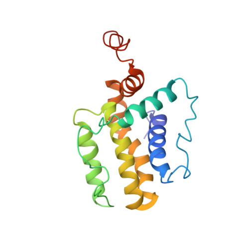
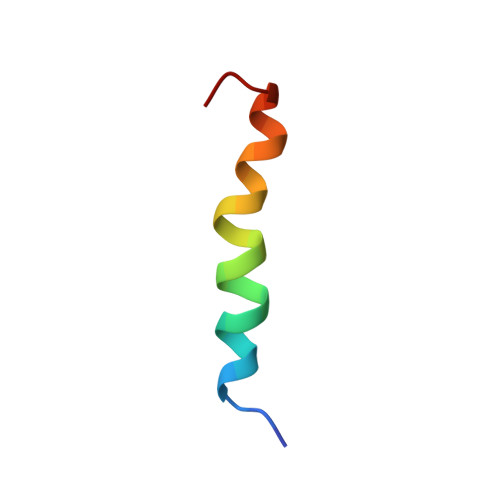
PUMA binding induces partial unfolding within BCL-xL to disrupt p53 binding and promote apoptosis.

Follis, A.V.Chipuk, J.E.Fisher, J.C.Yun, M.K.Grace, C.R.Nourse, A.Baran, K.Ou, L.Min, L.White, S.W.Green, D.R.Kriwacki, R.W.

(2013) Nat Chem Biol 9: 163-168

  • DOI: https://doi.org/10.1038/nchembio.1166
  • Primary Citation Related Structures: 
    2M03, 2M04, 4HNJ

  • PubMed Abstract: 

    Following DNA damage, nuclear p53 induces the expression of PUMA, a BH3-only protein that binds and inhibits the antiapoptotic BCL-2 repertoire, including BCL-xL. PUMA, unique among BH3-only proteins, disrupts the interaction between cytosolic p53 and BCL-xL, allowing p53 to promote apoptosis via direct activation of the BCL-2 effector molecules BAX and BAK. Structural investigations using NMR spectroscopy and X-ray crystallography revealed that PUMA binding induced partial unfolding of two α-helices within BCL-xL. Wild-type PUMA or a PUMA mutant incapable of causing binding-induced unfolding of BCL-xL equivalently inhibited the antiapoptotic BCL-2 repertoire to sensitize for death receptor-activated apoptosis, but only wild-type PUMA promoted p53-dependent, DNA damage-induced apoptosis. Our data suggest that PUMA-induced partial unfolding of BCL-xL disrupts interactions between cytosolic p53 and BCL-xL, releasing the bound p53 to initiate apoptosis. We propose that regulated unfolding of BCL-xL provides a mechanism to promote PUMA-dependent signaling within the apoptotic pathways.


  • Organizational Affiliation
    • Department of Structural Biology, St. Jude Children's Research Hospital, Memphis, Tennessee, USA.

Macromolecule Content 

  • Total Structure Weight: 23.73 kDa 
  • Atom Count: 1,612 
  • Modeled Residue Count: 199 
  • Deposited Residue Count: 205 
  • Unique protein chains: 2

Macromolecules

Find similar proteins by:|  3D Structure
Entity ID: 1
MoleculeChains  Sequence LengthOrganismDetailsImage
Bcl-2-like protein 1180Homo sapiensMutation(s): 0 
Gene Names: BCL2-like 1BCL2LBCL2L1BCLX
UniProt & NIH Common Fund Data Resources
Find proteins for Q07817 (Homo sapiens)
Explore Q07817 
Go to UniProtKB:  Q07817
PHAROS:  Q07817
GTEx:  ENSG00000171552 
Entity Groups
Sequence Clusters30% Identity50% Identity70% Identity90% Identity95% Identity100% Identity
UniProt GroupQ07817
Sequence Annotations
Expand
Reference Sequence
Find similar proteins by:  Sequence   |   3D Structure  
Entity ID: 2
MoleculeChains  Sequence LengthOrganismDetailsImage
Bcl-2-binding component 325Homo sapiensMutation(s): 0 
UniProt & NIH Common Fund Data Resources
Find proteins for Q9BXH1 (Homo sapiens)
Explore Q9BXH1 
Go to UniProtKB:  Q9BXH1
PHAROS:  Q9BXH1
GTEx:  ENSG00000105327 
Entity Groups
Sequence Clusters30% Identity50% Identity70% Identity90% Identity95% Identity100% Identity
UniProt GroupQ9BXH1
Sequence Annotations
Expand
Reference Sequence

Experimental Data & Validation

Experimental Data

  • Method: SOLUTION NMR
  • Conformers Calculated: 200 
  • Conformers Submitted: 20 
  • Selection Criteria: structures with the lowest energy 

Structure Validation

View Full Validation Report


Entry History 

Deposition Data

Revision History  (Full details and data files)

  • Version 1.0: 2013-01-30
    Type: Initial release
  • Version 1.1: 2013-02-06
    Changes: Database references
  • Version 1.2: 2013-03-06
    Changes: Database references
  • Version 1.3: 2024-05-01
    Changes: Data collection, Database references