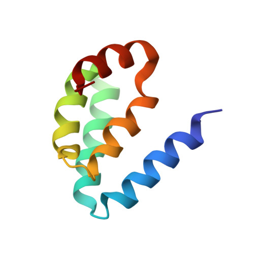
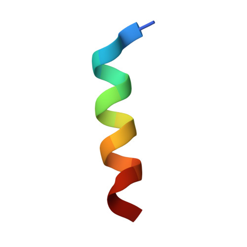
Large protein assemblies formed by multivalent interactions between cadherin23 and harmonin suggest a stable anchorage structure at the tip link of stereocilia.
Wu, L., Pan, L., Zhang, C., Zhang, M.(2012) J Biol Chem 287: 33460-33471
- PubMed: 22879593 Search on PubMedSearch on PubMed Central
- DOI: https://doi.org/10.1074/jbc.M112.378505
- Primary Citation Related Structures:
2LSR - PubMed Abstract:
Stereocilia tip links of inner ear hair cells are subjected to constant stretching during hair-bundle deflection, and accordingly are well designed to prevent from being broken by mechanical tensions. The roots of tip links, which couple tip links with the cytoskeleton, supposedly play important roles in withstanding large forces under stimulated conditions. The upper root of the tip link is mainly formed by the cytoplasmic tail of cadherin23 and its actin-anchoring protein harmonin. However, the detailed organization mode of the two proteins that gives rise to a strong upper root remains unclear. Here we show that the exon68-encoded peptide of cadherin23 can either interact with the N-terminal domain (NTD) of harmonin or form a homodimer. We demonstrate that the three harmonin binding sites of cadherin23, namely the NTD-binding motif, the exon68 peptide, and the C-terminal PDZ binding motif, do not synergize with each other in binding to harmonin, instead they facilitate formation of polymeric cadherin23/harmonin complexes. The exon68 peptide can promote the cadherin23/harmonin polymer formation via either binding to harmonin NTD or self-dimerization. We propose that the polymeric cadherin23/harmonin complex formed beneath the upper tip link membranes may serve as part of the stable rootlet structure for anchoring the tip links of stereocilia.
- Division of Life Science, State Key Laboratory of Molecular Neuroscience, Hong Kong University of Science and Technology, Clear Water Bay, Kowloon, Hong Kong.
Organizational Affiliation: