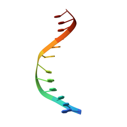
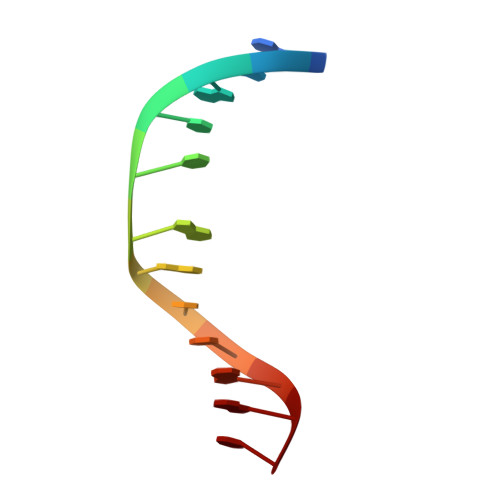
Conformational interconversion of the trans-4-hydroxynonenal-derived (6S,8R,11S) 1,N(2)-deoxyguanosine adduct when mismatched with deoxyadenosine in DNA
Huang, H., Wang, H., Lloyd, R.S., Rizzo, C.J., Stone, M.P.(2009) Chem Res Toxicol 22: 187-200
- PubMed: 19053179
- DOI: https://doi.org/10.1021/tx800320m
- Primary Citation Related Structures:
2KAR, 2KAS - PubMed Abstract:
The (6S,8R,11S) 1,N(2)-HNE-dGuo adduct of trans-4-hydroxynonenal (HNE) was incorporated into the duplex 5'-d(GCTAGCXAGTCC)-3'.5'-d(GGACTAGCTAGC)-3' [X = (6S,8R,11S) HNE-dG], in which the lesion was mismatched opposite dAdo. The (6S,8R,11S) adduct maintained the ring-closed 1,N(2)-HNE-dG structure. This was in contrast to when this adduct was correctly paired with dCyd, conditions under which it underwent ring opening and rearrangement to diastereomeric minor groove cyclic hemiacetals [ Huang , H. , Wang , H. , Qi , N. , Lloyd , R. S. , Harris , T. M. , Rizzo , C. J. , and Stone , M. P. ( 2008 ) J. Am. Chem. Soc. 130 , 10898 - 10906 ]. The (6S,8R,11S) adduct exhibited a syn/anti conformational equilibrium about the glycosyl bond. The syn conformation was predominant in acidic solution. Structural analysis of the syn conformation revealed that X(7) formed a distorted base pair with the complementary protonated A(18). The HNE moiety was located in the major groove. Structural perturbations were observed at the neighbor C(6).G(19) and A(8).T(17) base pairs. At basic pH, the anti conformation of X(7) was the major species. The 1,N(2)-HNE-dG intercalated and displaced the complementary A(18) in the 5'-direction, resulting in a bulge at the X(7).A(18) base pair. The HNE aliphatic chain was oriented toward the minor groove. The Watson-Crick hydrogen bonding of the neighboring A(8).T(17) base pair was also disrupted.
- Department of Chemistry, Center in Molecular Toxicology, Center for Structural Biology and Vanderbilt-Ingram Cancer Center, Vanderbilt University, Nashville, Tennessee 37235, USA.
Organizational Affiliation: