2K1Q | pdb_00002k1q

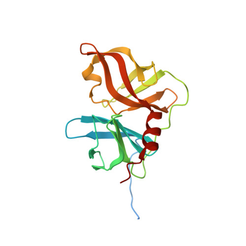
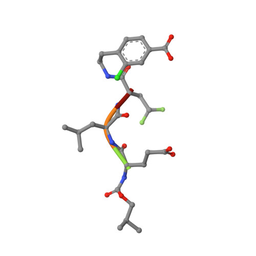
NMR structure of hepatitis c virus ns3 serine protease complexed with the non-covalently bound phenethylamide inhibitor


Experimental Data Snapshot

  • Method: SOLUTION NMR
  • Conformers Calculated: 200 
  • Conformers Submitted: 20 
  • Selection Criteria: structures with the lowest energy 

wwPDB Validation 3D Report Full Report

Validation slider image for 2K1Q

This is version 2.0 of the entry. See complete history. 

Literature

Binding of a noncovalent inhibitor exploiting the S' region stabilizes the hepatitis C virus NS3 protease conformation in the absence of cofactor.

Gallo, M., Pennestri, M., Bottomley, M.J., Barbato, G., Eliseo, T., Paci, M., Narjes, F., De Francesco, R., Summa, V., Koch, U., Bazzo, R., Cicero, D.O.

(2009) J Mol Biol 385: 1142-1155

  • DOI: https://doi.org/10.1016/j.jmb.2008.11.017
  • Primary Citation Related Structures: 
    2K1Q

  • PubMed Abstract: 

    We present the first structure of a noncovalent inhibitor bound to the protease domain of hepatitis C virus NS3 protein (NS3p), solved by NMR. The inhibitor exploits interactions with the S' region of NS3p to form a long-lived complex, although the absence of negative charges strongly reduces the association rate. The inhibitor stabilizes the N-terminal domain of NS3p and the substrate-binding site, and correctly aligns catalytic His-Asp residues. These actions were previously attributed exclusively to the cofactor NS4A, which interacts with the N-terminal domain of the NS3p and functions as an activator in vivo. The structure of the inhibitor/NS3p complex is very similar to that of the NS3p-NS4A complex, showing that binding of the NS4A cofactor is not the only event leading to a stable active-site conformation.


  • Organizational Affiliation: 
    • Department of Chemical Science and Technology, Università di Roma Tor Vergata Via della Ricerca Scientifica 1, 00133 Rome, Italy.

Macromolecule Content 

  • Total Structure Weight: 20.36 kDa 
  • Atom Count: 1,265 
  • Modeled Residue Count: 170 
  • Deposited Residue Count: 191 
  • Unique protein chains: 2

Macromolecules

Find similar proteins by:|  3D Structure
Entity ID: 1
MoleculeChains  Sequence LengthOrganismDetailsImage
NS3 PROTEASE186Orthohepacivirus hominisMutation(s): 0 
UniProt
Find proteins for P90191 (Orthohepacivirus hominis)
Explore P90191 
Go to UniProtKB:  P90191
Entity Groups
Sequence Clusters30% Identity50% Identity70% Identity90% Identity95% Identity100% Identity
UniProt GroupP90191
Sequence Annotations
Expand
Reference Sequence
Find similar proteins by:  Sequence   |   3D Structure  
Entity ID: 2
MoleculeChains  Sequence LengthOrganismDetailsImage
PHENETHYLAMIDE5N/AMutation(s): 0 
Sequence Annotations
Expand
Reference Sequence

Small Molecules

Ligands 1 Unique
IDChains Name / Formula / InChI Key2D Diagram3D Interactions
ZN

Query on ZN


Download:Ideal Coordinates CCD File
C [auth A]ZINC ION
Zn
PTFCDOFLOPIGGS-UHFFFAOYSA-N
Modified Residues  1 Unique
IDChains TypeFormula2D DiagramParent
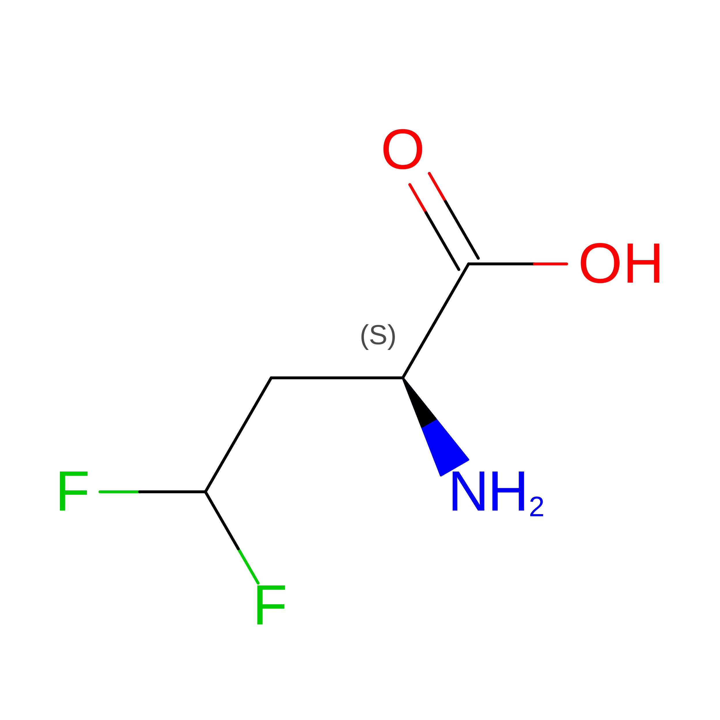
OBF
Query on OBF
B
L-PEPTIDE LINKINGC4 H7 F2 N O2

--

Experimental Data & Validation

Experimental Data

  • Method: SOLUTION NMR
  • Conformers Calculated: 200 
  • Conformers Submitted: 20 
  • Selection Criteria: structures with the lowest energy 

Structure Validation

View Full Validation Report


Entry History 

Deposition Data

Revision History  (Full details and data files)

  • Version 1.0: 2009-02-03
    Type: Initial release
  • Version 1.1: 2011-07-13
    Changes: Version format compliance
  • Version 1.2: 2022-03-16
    Changes: Data collection, Database references, Derived calculations
  • Version 2.0: 2023-11-15
    Changes: Atomic model, Data collection