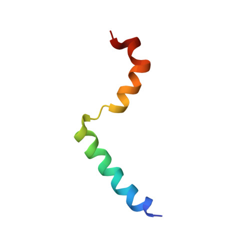
Three-dimensional structure of the two peptides that constitute the two-peptide bacteriocin lactococcin G
Rogne, P., Fimland, G., Nissen-Meyer, J., Kristiansen, P.E.(2008) Biochim Biophys Acta 1784: 543-554
- PubMed: 18187052
- DOI: https://doi.org/10.1016/j.bbapap.2007.12.002
- Primary Citation of Related Structures:
2JPJ, 2JPK, 2JPL, 2JPM - PubMed Abstract:
The three-dimensional structures of the two peptides, lactococcin G-alpha (LcnG-alpha; contains 39 residues) and lactococcin G-beta (LcnG-beta, contains 35 residues), that constitute the two-peptide bacteriocin lactococcin G (LcnG) have been determined by nuclear magnetic resonance (NMR) spectroscopy in the presence of DPC micelles and TFE. In DPC, LcnG-alpha has an N-terminal alpha-helix (residues 3-21) that contains a GxxxG helix-helix interaction motif (residues 7-11) and a less well defined C-terminal alpha-helix (residues 24-34), and in between (residues 18-22) there is a second somewhat flexible GxxxG-motif. Its structure in TFE was similar. In DPC, LcnG-beta has an N-terminal alpha-helix (residues 6-19). The region from residues 20 to 35, which also contains a flexible GxxxG-motif (residues 18-22), appeared to be fairly unstructured in DPC. In the presence of TFE, however, the region between and including residues 23 and 32 formed a well defined alpha-helix. The N-terminal helix between and including residues 6 and 19 seen in the presence of DPC, was broken at residues 8 and 9 in the presence of TFE. The N-terminal helices, both in LcnG-alpha and -beta, are amphiphilic. We postulate that LcnG-alpha and -beta have a parallel orientation and interact through helix-helix interactions involving the first GxxxG (residues 7-11) motif in LcnG-alpha and the one (residues 18-22) in LcnG-beta, and that they thus lie in a staggered fashion relative to each other.
- Department of Molecular Biosciences, University of Oslo, Pb 1041 Blindern, 0316 Oslo, Norway.
Organizational Affiliation:


