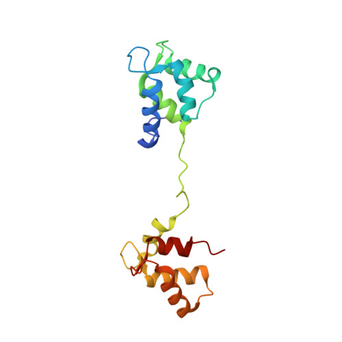
The structure of lethocerus troponin C: insights into the mechanism of stretch activation in muscles
De Nicola, G., Burkart, C., Qiu, F., Agianian, B., Labeit, S., Martin, S., Bullard, B., Pastore, A.(2007) Structure 15: 813-824
- PubMed: 17637342
- DOI: https://doi.org/10.1016/j.str.2007.05.007
- Primary Citation of Related Structures:
2JNF - PubMed Abstract:
To gain a molecular description of how muscles can be activated by mechanical stretch, we have solved the structure of the calcium-loaded F1 isoform of troponin C (TnC) from Lethocerus and characterized its interactions with troponin I (TnI). We show that the presence of only one calcium cation in the fourth EF hand motif is sufficient to induce an open conformation in the C-terminal lobe of F1 TnC, in contrast with what is observed in vertebrate muscle. This lobe interacts in a calcium-independent way both with the N terminus of TnI and, with lower affinity, with a region of TnI equivalent to the switch and inhibitory peptides of vertebrate muscles. Using both synthetic peptides and recombinant proteins, we show that the N lobe of F1 TnC is not engaged in interactions with TnI, excluding a regulatory role of this domain. These findings provide insights into mechanically stimulated muscle contraction.
- Molecular Structure Division, National Institute for Medical Research, The Ridgeway, Mill Hill, London NW7 1AA, United Kingdom.
Organizational Affiliation:


