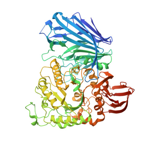
2JKP | pdb_00002jkp

Structure of a family 97 alpha-glucosidase from Bacteroides thetaiotaomicron in complex with castanospermine


Experimental Data Snapshot

  • Method: X-RAY DIFFRACTION
  • Resolution: 1.99 Å
  • R-Value Free: 
    0.227 (Depositor), 0.230 (DCC) 
  • R-Value Work: 
    0.173 (Depositor), 0.170 (DCC) 
  • R-Value Observed: 
    0.176 (Depositor) 

Starting Model: experimental
View more details

wwPDB Validation   3D Report Full Report


Ligand Structure Quality Assessment 

Created with Raphaël 2.3.0Worse 01 BetterLigand structure goodness of fit to experimental dataBest fitted CTSClick on this verticalbar to view details

This is version 1.3 of the entry. See complete history


Literature

Divergence of Catalytic Mechanism within a Glycosidase Family Provides Insight Into Evolution of Carbohydrate Metabolism by Human Gut Flora.

Gloster, T.M.Turkenburg, J.P.Potts, J.R.Henrissat, B.Davies, G.J.

(2008) Chem Biol 15: 1058

  • DOI: https://doi.org/10.1016/j.chembiol.2008.09.005
  • Primary Citation of Related Structures:  
    2JKA, 2JKE, 2JKP

  • PubMed Abstract: 

    Enzymatic cleavage of the glycosidic bond yields products in which the anomeric configuration is either retained or inverted. Each mechanism reflects the dispositions of the enzyme functional groups; a facet of which is essentially conserved in 113 glycoside hydrolase (GH) families. We show that family GH97 has diverged significantly, as it contains both inverting and retaining alpha-glycosidases. This reflects evolution of the active center; a glutamate acts as a general base in inverting members, exemplified by Bacteroides thetaiotaomicron alpha-glucosidase BtGH97a, whereas an aspartate likely acts as a nucleophile in retaining members. The structure of BtGH97a and its complexes with inhibitors, coupled to kinetic analysis of active-site variants, reveals an unusual calcium ion dependence. 1H NMR analysis shows an inversion mechanism for BtGH97a, whereas another GH97 enzyme from B. thetaiotaomicron, BtGH97b, functions as a retaining alpha-galactosidase.


  • Organizational Affiliation

    York Structural Biology Laboratory, Department of Chemistry, University of York, Heslington, York, YO10 5YW, UK. gloster@ysbl.york.ac.uk


Macromolecules
Find similar proteins by:  (by identity cutoff)  |  3D Structure
Entity ID: 1
MoleculeChains Sequence LengthOrganismDetailsImage
ALPHA-GLUCOSIDASE (ALPHA-GLUCOSIDASE SUSB)
A, B
727Bacteroides thetaiotaomicron VPI-5482Mutation(s): 0 
EC: 3.2.1.20 (PDB Primary Data), 3.2.1.3 (UniProt)
UniProt
Find proteins for G8JZS4 (Bacteroides thetaiotaomicron (strain ATCC 29148 / DSM 2079 / JCM 5827 / CCUG 10774 / NCTC 10582 / VPI-5482 / E50))
Explore G8JZS4 
Go to UniProtKB:  G8JZS4
Entity Groups  
Sequence Clusters30% Identity50% Identity70% Identity90% Identity95% Identity100% Identity
UniProt GroupG8JZS4
Sequence Annotations
Expand
  • Reference Sequence
Small Molecules
Ligands 3 Unique
IDChains Name / Formula / InChI Key2D Diagram3D Interactions
CTS
Query on CTS

Download Ideal Coordinates CCD File 
C [auth A],
G [auth B]
CASTANOSPERMINE
C8 H15 N O4
JDVVGAQPNNXQDW-TVNFTVLESA-N
EDO
Query on EDO

Download Ideal Coordinates CCD File 
E [auth A],
F [auth A],
I [auth B],
J [auth B]
1,2-ETHANEDIOL
C2 H6 O2
LYCAIKOWRPUZTN-UHFFFAOYSA-N
CA
Query on CA

Download Ideal Coordinates CCD File 
D [auth A],
H [auth B]
CALCIUM ION
Ca
BHPQYMZQTOCNFJ-UHFFFAOYSA-N
Experimental Data & Validation

Experimental Data

  • Method: X-RAY DIFFRACTION
  • Resolution: 1.99 Å
  • R-Value Free:  0.227 (Depositor), 0.230 (DCC) 
  • R-Value Work:  0.173 (Depositor), 0.170 (DCC) 
  • R-Value Observed: 0.176 (Depositor) 
Space Group: P 1 21 1
Unit Cell:
Length ( Å )Angle ( ˚ )
a = 74.566α = 90
b = 111.988β = 100.89
c = 104.134γ = 90
Software Package:
Software NamePurpose
REFMACrefinement
DENZOdata reduction
SCALEPACKdata scaling
REFMACphasing

Structure Validation

View Full Validation Report


Ligand Structure Quality Assessment 

Created with Raphaël 2.3.0Worse 01 BetterLigand structure goodness of fit to experimental dataBest fitted CTSClick on this verticalbar to view details

Entry History 

Deposition Data

Revision History  (Full details and data files)

  • Version 1.0: 2008-09-30
    Type: Initial release
  • Version 1.1: 2011-05-07
    Changes: Version format compliance
  • Version 1.2: 2011-07-13
    Changes: Version format compliance
  • Version 1.3: 2023-12-13
    Changes: Data collection, Database references, Derived calculations, Other, Refinement description