2JEY

Mus musculus acetylcholinesterase in complex with HLo-7


Experimental Data Snapshot

  • Method: X-RAY DIFFRACTION
  • Resolution: 2.70 Å
  • R-Value Free: 0.261 
  • R-Value Work: 0.206 
  • R-Value Observed: 0.207 

wwPDB Validation   3D Report Full Report


Ligand Structure Quality Assessment 


This is version 1.5 of the entry. See complete history


Literature

Novel Nerve-Agent Antidote Design Based on Crystallographic and Mass Spectrometric Analyses of Tabun-Conjugated Acetylcholinesterase in Complex with Antidotes.

Ekstrom, F.J.Astot, C.Pang, Y.

(2007) Clin Pharmacol Ther 82: 282

  • DOI: https://doi.org/10.1038/sj.clpt.6100151
  • Primary Citation of Related Structures:  
    2JEY, 2JEZ, 2JF0

  • PubMed Abstract: 

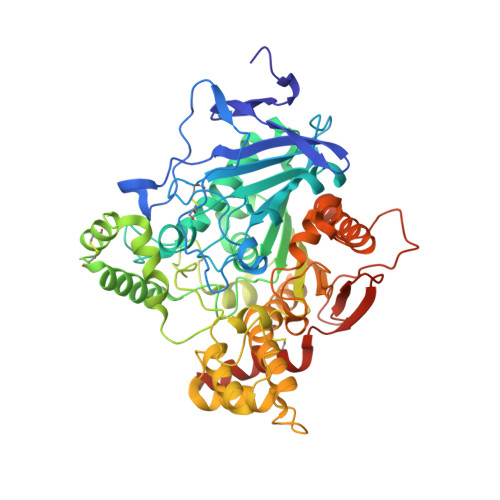
    Organophosphorus compound-based nerve agents inhibit the essential enzyme acetylcholinesterase (AChE) causing acute toxicity and death. Clinical treatment of nerve-agent poisoning is to use oxime-based antidotes to reactivate the inhibited AChE. However, the nerve agent tabun is resistant to oximes. To design improved oximes, crystal structures of a tabun-conjugated AChE in complex with different oximes are needed to guide the structural modifications of known antidotes. However, this type of structure is extremely challenging to obtain because both deamidation of the tabun conjugate and reactivation of AChE occur during crystallographic experiments. Here we report, for the first time, the crystal structures of Ortho-7 and HLö-7 in complex with AChE that is conjugated to an intact tabun. These structures were determined by our new strategy of combining crystallographic and mass spectrometric analyses of AChE crystals. The results explain the relative reactivation potencies of the two oximes and offer insights into improving known medical antidotes.


  • Organizational Affiliation

    FOI CBRN Defence and Security, Umeå, Sweden. fredrik.ekstrom@foi.se


Macromolecules
Find similar proteins by:  (by identity cutoff)  |  3D Structure
Entity ID: 1
MoleculeChains Sequence LengthOrganismDetailsImage
ACETYLCHOLINESTERASE
A, B
548Mus musculusMutation(s): 0 
EC: 3.1.1.7 (PDB Primary Data), 3.1.1.1 (PDB Primary Data)
UniProt & NIH Common Fund Data Resources
Find proteins for P21836 (Mus musculus)
Explore P21836 
Go to UniProtKB:  P21836
IMPC:  MGI:87876
Entity Groups  
Sequence Clusters30% Identity50% Identity70% Identity90% Identity95% Identity100% Identity
UniProt GroupP21836
Sequence Annotations
Expand
  • Reference Sequence
Small Molecules
Ligands 2 Unique
IDChains Name / Formula / InChI Key2D Diagram3D Interactions
HLO
Query on HLO

Download Ideal Coordinates CCD File 
C [auth A],
D [auth B]
1-[({2,4-BIS[(E)-(HYDROXYIMINO)METHYL]PYRIDINIUM-1-YL}METHOXY)METHYL]-4-CARBAMOYLPYRIDINIUM
C15 H17 N5 O4
SODKCPHRJDOHMI-UHFFFAOYSA-P
P6G
Query on P6G

Download Ideal Coordinates CCD File 
E [auth B]HEXAETHYLENE GLYCOL
C12 H26 O7
IIRDTKBZINWQAW-UHFFFAOYSA-N
Experimental Data & Validation

Experimental Data

  • Method: X-RAY DIFFRACTION
  • Resolution: 2.70 Å
  • R-Value Free: 0.261 
  • R-Value Work: 0.206 
  • R-Value Observed: 0.207 
  • Space Group: P 21 21 21
Unit Cell:
Length ( Å )Angle ( ˚ )
a = 76.67α = 90
b = 108.45β = 90
c = 220.62γ = 90
Software Package:
Software NamePurpose
REFMACrefinement
XDSdata reduction
SCALAdata scaling
REFMACphasing

Structure Validation

View Full Validation Report


Ligand Structure Quality Assessment 


Entry History 

Deposition Data

Revision History  (Full details and data files)

  • Version 1.0: 2007-07-03
    Type: Initial release
  • Version 1.1: 2011-05-08
    Changes: Version format compliance
  • Version 1.2: 2011-07-13
    Changes: Version format compliance
  • Version 1.3: 2018-01-17
    Changes: Data collection
  • Version 1.4: 2021-05-12
    Changes: Derived calculations, Other
  • Version 1.5: 2023-12-13
    Changes: Data collection, Database references, Refinement description