2JEU | pdb_00002jeu

Transcription activator structure reveals redox control of a replication initiation reaction


Experimental Data Snapshot

  • Method: X-RAY DIFFRACTION
  • Resolution: 2.80 Å
  • R-Value Free: 
    0.281 (Depositor) 
  • R-Value Work: 
    0.233 (Depositor), 0.234 (DCC) 
  • R-Value Observed: 
    0.236 (Depositor) 

Starting Model: experimental
View more details

wwPDB Validation 3D Report Full Report

Validation slider image for 2JEU

This is version 1.3 of the entry. See complete history

Literature

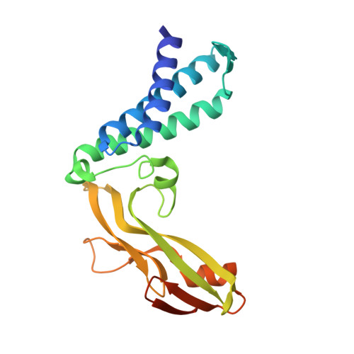
Transcription Activator Structure Reveals Redox Control of a Replication Initiation Reaction.

Sanders, C.M.Sizov, D.Seavers, P.R.Ortiz-Lombardia, M.Antson, A.A.

(2007) Nucleic Acids Res 35: 3504

  • DOI: https://doi.org/10.1093/nar/gkm166
  • Primary Citation Related Structures: 
    2JEU, 2JEX

  • PubMed Abstract: 

    Redox changes are one of the factors that influence cell-cycle progression and that control the processes of cellular proliferation, differentiation, senescence and apoptosis. Proteins regulated through redox-sensitive cysteines have been characterized but specific 'sulphydryl switches' in replication proteins remain to be identified. In bovine papillomavirus type-1, DNA replication begins when the viral transcription factor E2 recruits the viral initiator protein E1 to the origin of DNA replication (ori). Here we show that a novel dimerization interface in the E2 transcription activation domain is stabilized by a disulphide bond. Oxidative cross-linking via Cys57 sequesters the interaction surface between E1 and E2, preventing pre-initiation and replication initiation complex formation. Our data demonstrate that as well as a mechanism for regulating DNA binding, redox reactions can control replication by modulating the tertiary structure of critical protein factors using a specific redox sensor.


  • Organizational Affiliation
    • Institute for Cancer Studies, University of Sheffield, Beech Hill Rd, Sheffield, S10 2RX, UK. c.m.sanders@sheffield.ac.uk

Macromolecule Content 

  • Total Structure Weight: 23.91 kDa 
  • Atom Count: 1,595 
  • Modeled Residue Count: 197 
  • Deposited Residue Count: 209 
  • Unique protein chains: 1

Macromolecules

Find similar proteins by:|  3D Structure
Entity ID: 1
MoleculeChains  Sequence LengthOrganismDetailsImage
REGULATORY PROTEIN E2209Deltapapillomavirus 4Mutation(s): 0 
UniProt
Find proteins for P03122 (Bovine papillomavirus type 1)
Explore P03122 
Go to UniProtKB:  P03122
Entity Groups
Sequence Clusters30% Identity50% Identity70% Identity90% Identity95% Identity100% Identity
UniProt GroupP03122
Sequence Annotations
Expand
Reference Sequence

Experimental Data & Validation

Experimental Data

  • Method: X-RAY DIFFRACTION
  • Resolution: 2.80 Å
  • R-Value Free:  0.281 (Depositor) 
  • R-Value Work:  0.233 (Depositor), 0.234 (DCC) 
  • R-Value Observed: 0.236 (Depositor) 
Space Group: P 61 2 2
Unit Cell:
Length ( Å )Angle ( ˚ )
a = 61.539α = 90
b = 61.539β = 90
c = 236.317γ = 120
Software Package:
Software NamePurpose
REFMACrefinement
DENZOdata reduction
SCALEPACKdata scaling
AMoREphasing

Structure Validation

View Full Validation Report


Entry History 

Deposition Data

Revision History  (Full details and data files)

  • Version 1.0: 2007-05-15
    Type: Initial release
  • Version 1.1: 2011-07-13
    Changes: Advisory, Version format compliance
  • Version 1.2: 2019-05-08
    Changes: Data collection, Experimental preparation, Other
  • Version 1.3: 2023-12-13
    Changes: Data collection, Database references, Other, Refinement description