2JE1

The crystal Structure of the tumor supressor protein pp32 (Anp32a) :structural insights into the Anp32 family of proteins


Experimental Data Snapshot

  • Method: X-RAY DIFFRACTION
  • Resolution: 2.69 Å
  • R-Value Free: 0.303 
  • R-Value Work: 0.238 
  • R-Value Observed: 0.241 

wwPDB Validation   3D Report Full Report


This is version 1.1 of the entry. See complete history


Literature

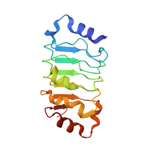
The Crystal Structure of the Tumor Suppressor Protein Pp32 (Anp32A): Structural Insights Into Anp32 Family of Proteins.

Huyton, T.Wolberger, C.

(2007) Protein Sci 16: 1308

  • DOI: https://doi.org/10.1110/ps.072803507
  • Primary Citation of Related Structures:  
    2JE0, 2JE1

  • PubMed Abstract: 

    The tumor suppressor protein pp32 is highly overexpressed in many cancers of the breast and prostate, and has also been implicated in the neurodegenerative disease spinocerebellar ataxias type 1 (SCA1). Pp32 is a multifunctional protein that is involved in the regulation of transcription, apoptosis, phosphorylation, and cell cycle progression, the latter through its association with the hyperphosphorylated form of the retinoblastoma tumor suppressor. We have determined the structure of an N-terminal pp32 fragment comprising a capped leucine-rich repeat (LRR) domain, which provides insight into the structural and biochemical properties of the pp32 (Anp32) family of proteins.


  • Organizational Affiliation

    Department of Biophysics and Biophysical Chemistry, Howard Hughes Medical Institute, Johns Hopkins University School of Medicine, Baltimore, MD 21205-2185, USA.


Macromolecules
Find similar proteins by:  (by identity cutoff)  |  3D Structure
Entity ID: 1
MoleculeChains Sequence LengthOrganismDetailsImage
ACIDIC LEUCINE-RICH NUCLEAR PHOSPHOPROTEIN 32 FAMILY MEMBER A
A, B, C, D
149Homo sapiensMutation(s): 0 
UniProt & NIH Common Fund Data Resources
Find proteins for P39687 (Homo sapiens)
Explore P39687 
Go to UniProtKB:  P39687
PHAROS:  P39687
GTEx:  ENSG00000140350 
Entity Groups  
Sequence Clusters30% Identity50% Identity70% Identity90% Identity95% Identity100% Identity
UniProt GroupP39687
Sequence Annotations
Expand
  • Reference Sequence
Small Molecules
Ligands 1 Unique
IDChains Name / Formula / InChI Key2D Diagram3D Interactions
GOL
Query on GOL

Download Ideal Coordinates CCD File 
E [auth A]GLYCEROL
C3 H8 O3
PEDCQBHIVMGVHV-UHFFFAOYSA-N
Experimental Data & Validation

Experimental Data

  • Method: X-RAY DIFFRACTION
  • Resolution: 2.69 Å
  • R-Value Free: 0.303 
  • R-Value Work: 0.238 
  • R-Value Observed: 0.241 
  • Space Group: P 63
Unit Cell:
Length ( Å )Angle ( ˚ )
a = 106.506α = 90
b = 106.506β = 90
c = 133.153γ = 120
Software Package:
Software NamePurpose
REFMACrefinement
DENZOdata reduction
SCALEPACKdata scaling
MOLREPphasing

Structure Validation

View Full Validation Report


Entry History 

Deposition Data

Revision History  (Full details and data files)

  • Version 1.0: 2007-06-26
    Type: Initial release
  • Version 1.1: 2011-07-13
    Changes: Advisory, Version format compliance