2J94

CRYSTAL STRUCTURE OF A HUMAN FACTOR XA INHIBITOR COMPLEX


Experimental Data Snapshot

  • Method: X-RAY DIFFRACTION
  • Resolution: 2.10 Å
  • R-Value Free: 0.233 
  • R-Value Work: 0.181 
  • R-Value Observed: 0.184 

wwPDB Validation   3D Report Full Report


Ligand Structure Quality Assessment 


This is version 1.3 of the entry. See complete history


Literature

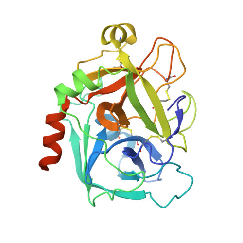
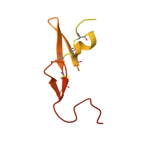
Factor Xa Inhibitors: S1 Binding Interactions of a Series of N-{(3S)-1-[(1S)-1-Methyl-2-Morpholin-4-Yl-2-Oxoethyl]-2-Oxopyrrolidin-3-Yl}Sulfonamides.

Chan, C.Borthwick, A.D.Brown, D.Burns-Kurtis, C.L.Campbell, M.Chaudry, L.Chung, C.W.Convery, M.A.Hamblin, J.N.Johnstone, L.Kelly, H.A.Kleanthous, S.Patikis, A.Patel, C.Pateman, A.J.Senger, S.Shah, G.P.Toomey, J.R.Watson, N.S.Weston, H.E.Whitworth, C.Young, R.J.Zhou, P.

(2007) J Med Chem 50: 1546

  • DOI: https://doi.org/10.1021/jm060870c
  • Primary Citation of Related Structures:  
    2J94, 2J95, 4Y76, 4Y79

  • PubMed Abstract: 

    Factor Xa inhibitory activities for a series of N-{(3S)-1-[(1S)-1-methyl-2-morpholin-4-yl-2-oxoethyl]-2-oxopyrrolidin-3-yl}sulfonamides with different P1 groups are described. These data provide insight into binding interactions within the S1 primary specificity pocket; rationales are presented for the derived SAR on the basis of electronic interactions through crystal structures of fXa-ligand complexes and molecular modeling studies. A good correlation between in vitro anticoagulant activities with lipophilicity and the extent of human serum albumin binding is observed within this series of potent fXa inhibitors. Pharmacokinetic profiles in rat and dog, together with selectivity over other trypsin-like serine proteases, identified 1f as a candidate for further evaluation.


  • Organizational Affiliation

    CVU UK Medicinal Chemistry Department, GlaxoSmithKline Research and Development, Medicines Research Centre, Gunnels Wood Road, Stevenage, Hertfordshire SG1 2NY, United Kingdom. chuen.8chan@gsk.com


Macromolecules
Find similar proteins by:  (by identity cutoff)  |  3D Structure
Entity ID: 1
MoleculeChains Sequence LengthOrganismDetailsImage
COAGULATION FACTOR X254Homo sapiensMutation(s): 0 
EC: 3.4.21.6
UniProt & NIH Common Fund Data Resources
Find proteins for P00742 (Homo sapiens)
Explore P00742 
Go to UniProtKB:  P00742
PHAROS:  P00742
GTEx:  ENSG00000126218 
Entity Groups  
Sequence Clusters30% Identity50% Identity70% Identity90% Identity95% Identity100% Identity
UniProt GroupP00742
Sequence Annotations
Expand
  • Reference Sequence
Find similar proteins by:  (by identity cutoff)  |  3D Structure
Entity ID: 2
MoleculeChains Sequence LengthOrganismDetailsImage
COAGULATION FACTOR X134Homo sapiensMutation(s): 0 
EC: 3.4.21.6
UniProt & NIH Common Fund Data Resources
Find proteins for P00742 (Homo sapiens)
Explore P00742 
Go to UniProtKB:  P00742
PHAROS:  P00742
GTEx:  ENSG00000126218 
Entity Groups  
Sequence Clusters30% Identity50% Identity70% Identity90% Identity95% Identity100% Identity
UniProt GroupP00742
Sequence Annotations
Expand
  • Reference Sequence
Small Molecules
Ligands 2 Unique
IDChains Name / Formula / InChI Key2D Diagram3D Interactions
G15
Query on G15

Download Ideal Coordinates CCD File 
C [auth A]5-(5-CHLORO-2-THIENYL)-N-{(3S)-1-[(1S)-1-METHYL-2-MORPHOLIN-4-YL-2-OXOETHYL]-2-OXOPYRROLIDIN-3-YL}-1H-1,2,4-TRIAZOLE-3-SULFONAMIDE
C17 H21 Cl N6 O5 S2
FKCQUVAGEOSYRU-QWRGUYRKSA-N
CA
Query on CA

Download Ideal Coordinates CCD File 
D [auth A]CALCIUM ION
Ca
BHPQYMZQTOCNFJ-UHFFFAOYSA-N
Binding Affinity Annotations 
IDSourceBinding Affinity
G15 PDBBind:  2J94 Ki: 534 (nM) from 1 assay(s)
Binding MOAD:  2J94 Ki: 534 (nM) from 1 assay(s)
Experimental Data & Validation

Experimental Data

  • Method: X-RAY DIFFRACTION
  • Resolution: 2.10 Å
  • R-Value Free: 0.233 
  • R-Value Work: 0.181 
  • R-Value Observed: 0.184 
  • Space Group: P 21 21 21
Unit Cell:
Length ( Å )Angle ( ˚ )
a = 56.739α = 90
b = 72.563β = 90
c = 78.924γ = 90
Software Package:
Software NamePurpose
REFMACrefinement
MOSFLMdata reduction
SCALAdata scaling

Structure Validation

View Full Validation Report


Ligand Structure Quality Assessment 


Entry History 

Revision History  (Full details and data files)

  • Version 1.0: 2007-03-20
    Type: Initial release
  • Version 1.1: 2011-05-08
    Changes: Version format compliance
  • Version 1.2: 2011-07-13
    Changes: Version format compliance
  • Version 1.3: 2023-12-13
    Changes: Data collection, Database references, Derived calculations, Other, Refinement description