2J8W

The crystal structure of cytochrome c' from Rubrivivax gelatinosus at 1.3 A Resolution and pH 8.0


Experimental Data Snapshot

  • Method: X-RAY DIFFRACTION
  • Resolution: 1.29 Å
  • R-Value Free: 0.179 
  • R-Value Work: 0.151 
  • R-Value Observed: 0.151 

wwPDB Validation   3D Report Full Report


Ligand Structure Quality Assessment 


This is version 2.4 of the entry. See complete history


Literature

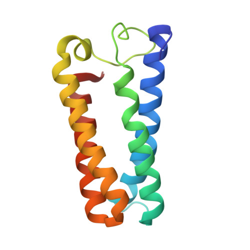
High resolution crystal structure of Rubrivivax gelatinosus cytochrome c'.

Benini, S.Rypniewski, W.R.Wilson, K.S.Ciurli, S.

(2008) J Inorg Biochem 102: 1322-1328

  • DOI: https://doi.org/10.1016/j.jinorgbio.2008.01.017
  • Primary Citation of Related Structures:  
    2J8W, 2J9B

  • PubMed Abstract: 

    The structure of the cytochrome c' from the purple non-sulfur phototrophic bacterium Rubrivivax gelatinosus was determined using two crystals grown independently at pH 6.3 and pH 8. The resolution attained for the two structures (1.29 A and 1.50 A for the crystals at high and low pH, respectively) is the highest to date for this class of proteins. The two structures were compared in detail in an attempt to investigate the influence of pH on the geometry of the haem and of the coordination environment of the Fe(III) ion. However, while the results suggest some small propensity for the movement of the metal atom out of the plane of the haem ring upon pH increase, the accuracy of the measurements at these two pH below the pK of the axial histidine is not sufficient to provide hard evidence of a shift in the iron position and associated changes.


  • Organizational Affiliation

    York Structural Biology Laboratory, Department of Chemistry, University of York, Heslington, York, YO10 5YW, United Kingdom.


Macromolecules
Find similar proteins by:  (by identity cutoff)  |  3D Structure
Entity ID: 1
MoleculeChains Sequence LengthOrganismDetailsImage
CYTOCHROME C'
A, B
129Rubrivivax gelatinosusMutation(s): 0 
UniProt
Find proteins for P00142 (Rubrivivax gelatinosus)
Explore P00142 
Go to UniProtKB:  P00142
Entity Groups  
Sequence Clusters30% Identity50% Identity70% Identity90% Identity95% Identity100% Identity
UniProt GroupP00142
Sequence Annotations
Expand
  • Reference Sequence
Experimental Data & Validation

Experimental Data

  • Method: X-RAY DIFFRACTION
  • Resolution: 1.29 Å
  • R-Value Free: 0.179 
  • R-Value Work: 0.151 
  • R-Value Observed: 0.151 
  • Space Group: P 31 2 1
Unit Cell:
Length ( Å )Angle ( ˚ )
a = 69.63α = 90
b = 69.63β = 90
c = 123.63γ = 120
Software Package:
Software NamePurpose
REFMACrefinement
DENZOdata reduction
SCALEPACKdata scaling
AMoREphasing

Structure Validation

View Full Validation Report


Ligand Structure Quality Assessment 


Entry History 

Deposition Data

Revision History  (Full details and data files)

  • Version 1.0: 2007-11-06
    Type: Initial release
  • Version 1.1: 2011-05-08
    Changes: Version format compliance
  • Version 1.2: 2011-07-13
    Changes: Version format compliance
  • Version 2.0: 2019-01-23
    Changes: Atomic model, Data collection, Database references, Derived calculations, Non-polymer description, Structure summary
  • Version 2.1: 2019-05-08
    Changes: Data collection, Derived calculations, Experimental preparation
  • Version 2.2: 2019-07-10
    Changes: Data collection
  • Version 2.3: 2019-07-24
    Changes: Data collection
  • Version 2.4: 2023-12-13
    Changes: Data collection, Database references, Derived calculations, Other, Refinement description