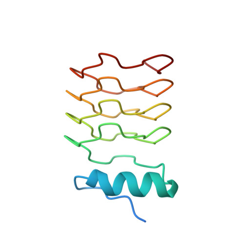
2J8I

Structure of NP275, a pentapeptide repeat protein from Nostoc punctiforme


Experimental Data Snapshot

  • Method: X-RAY DIFFRACTION
  • Resolution: 2.10 Å
  • R-Value Free: 0.256 
  • R-Value Work: 0.188 
  • R-Value Observed: 0.191 

Starting Model: experimental
View more details

wwPDB Validation   3D Report Full Report


This is version 1.2 of the entry. See complete history


Literature

Structural Characterization of the Fusion of Two Pentapeptide Repeat Proteins, Np275 and Np276, from Nostoc Punctiforme: Resurrection of an Ancestral Protein.

Vetting, M.W.Hegde, S.S.Hazleton, K.Z.Blanchard, J.S.

(2007) Protein Sci 16: 755

  • DOI: https://doi.org/10.1110/ps.062637707
  • Primary Citation of Related Structures:  
    2J8I, 2J8K

  • PubMed Abstract: 

    The Nostoc punctiforme genes Np275 and Np276 are two adjacently encoded proteins of 98 and 75 amino acids in length and exhibit sequences composed of tandem pentapeptide repeats. The structures of Np275 and a fusion of Np275 and Np276 were determined to 2.1 and 1.5 A, respectively. The two Nostoc proteins fold as highly symmetric right-handed quadrilateral beta-helices similar to the mycobacterial protein MfpA implicated in fluoroquinolone resistance and DNA gyrase inhibition. The sequence composition of the intervening coding region and the ability to express a fused protein by removing the stop codon for Np275 suggests Np275 and Np276 were recently part of a larger ancestral pentapeptide repeat protein.


  • Organizational Affiliation

    Department of Biochemistry, Albert Einstein College of Medicine, Bronx, New York 10461, USA.


Macromolecules
Find similar proteins by:  (by identity cutoff)  |  3D Structure
Entity ID: 1
MoleculeChains Sequence LengthOrganismDetailsImage
NP275
A, B
118Nostoc punctiformeMutation(s): 0 
Entity Groups  
Sequence Clusters30% Identity50% Identity70% Identity90% Identity95% Identity100% Identity
Sequence Annotations
Expand
  • Reference Sequence
Small Molecules
Ligands 1 Unique
IDChains Name / Formula / InChI Key2D Diagram3D Interactions
CL
Query on CL

Download Ideal Coordinates CCD File 
C [auth B]CHLORIDE ION
Cl
VEXZGXHMUGYJMC-UHFFFAOYSA-M
Experimental Data & Validation

Experimental Data

  • Method: X-RAY DIFFRACTION
  • Resolution: 2.10 Å
  • R-Value Free: 0.256 
  • R-Value Work: 0.188 
  • R-Value Observed: 0.191 
  • Space Group: P 21 21 21
Unit Cell:
Length ( Å )Angle ( ˚ )
a = 29.181α = 90
b = 62.558β = 90
c = 100.549γ = 90
Software Package:
Software NamePurpose
REFMACrefinement
MOSFLMdata reduction
CCP4data scaling
MOLREPphasing

Structure Validation

View Full Validation Report


Entry History 

Deposition Data

Revision History  (Full details and data files)

  • Version 1.0: 2006-12-07
    Type: Initial release
  • Version 1.1: 2011-07-13
    Changes: Advisory, Version format compliance
  • Version 1.2: 2023-12-13
    Changes: Data collection, Database references, Other, Refinement description