2J55

X-ray reduced Paraccocus denitrificans methylamine dehydrogenase O- quinone in complex with amicyanin.


Experimental Data Snapshot

  • Method: X-RAY DIFFRACTION
  • Resolution: 2.15 Å
  • R-Value Free: 0.245 
  • R-Value Work: 0.189 
  • R-Value Observed: 0.192 

Starting Model: other
View more details

wwPDB Validation   3D Report Full Report


This is version 1.2 of the entry. See complete history


Literature

Tracking X-Ray-Derived Redox Changes in Crystals of a Methylamine Dehydrogenase/Amicyanin Complex Using Single-Crystal Uv/Vis Microspectrophotometry.

Pearson, A.R.Pahl, R.Kovaleva, E.G.Davidson, V.L.Wilmot, C.M.

(2007) J Synchrotron Radiat 14: 92

  • DOI: https://doi.org/10.1107/S0909049506051259
  • Primary Citation of Related Structures:  
    2J55, 2J56, 2J57

  • PubMed Abstract: 

    X-ray exposure during crystallographic data collection can result in unintended redox changes in proteins containing functionally important redox centers. In order to directly monitor X-ray-derived redox changes in trapped oxidative half-reaction intermediates of Paracoccus denitrificans methylamine dehydrogenase, a commercially available single-crystal UV/Vis microspectrophotometer was installed on-line at the BioCARS beamline 14-BM-C at the Advanced Photon Source, Argonne, USA. Monitoring the redox state of the intermediates during X-ray exposure permitted the creation of a general multi-crystal data collection strategy to generate true structures of each redox intermediate.


  • Organizational Affiliation

    Department of Biochemistry, Molecular Biology and Biophysics, The University of Minnesota, Minneapolis, MN 55455, USA.


Macromolecules
Find similar proteins by:  (by identity cutoff)  |  3D Structure
Entity ID: 1
MoleculeChains Sequence LengthOrganismDetailsImage
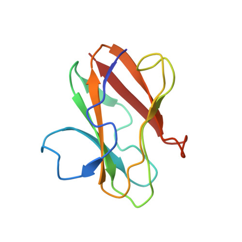
AMICYANIN
A, B
105Paracoccus denitrificansMutation(s): 0 
UniProt
Find proteins for P22364 (Paracoccus denitrificans)
Explore P22364 
Go to UniProtKB:  P22364
Entity Groups  
Sequence Clusters30% Identity50% Identity70% Identity90% Identity95% Identity100% Identity
UniProt GroupP22364
Sequence Annotations
Expand
  • Reference Sequence
Find similar proteins by:  (by identity cutoff)  |  3D Structure
Entity ID: 2
MoleculeChains Sequence LengthOrganismDetailsImage
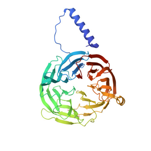
METHYLAMINE DEHYDROGENASE HEAVY CHAINC [auth H],
D [auth J]
386Paracoccus denitrificansMutation(s): 0 
EC: 1.4.99.3
UniProt
Find proteins for P29894 (Paracoccus denitrificans)
Explore P29894 
Go to UniProtKB:  P29894
Entity Groups  
Sequence Clusters30% Identity50% Identity70% Identity90% Identity95% Identity100% Identity
UniProt GroupP29894
Sequence Annotations
Expand
  • Reference Sequence
Find similar proteins by:  (by identity cutoff)  |  3D Structure
Entity ID: 3
MoleculeChains Sequence LengthOrganismDetailsImage
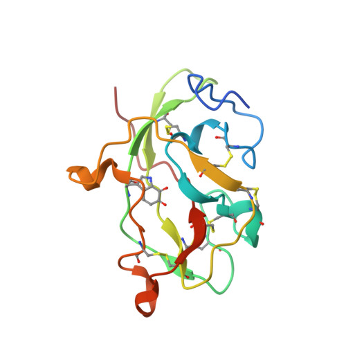
METHYLAMINE DEHYDROGENASE LIGHT CHAINE [auth L],
F [auth M]
131Paracoccus denitrificansMutation(s): 0 
EC: 1.4.99.3
UniProt
Find proteins for P22619 (Paracoccus denitrificans)
Explore P22619 
Go to UniProtKB:  P22619
Entity Groups  
Sequence Clusters30% Identity50% Identity70% Identity90% Identity95% Identity100% Identity
UniProt GroupP22619
Sequence Annotations
Expand
  • Reference Sequence
Experimental Data & Validation

Experimental Data

  • Method: X-RAY DIFFRACTION
  • Resolution: 2.15 Å
  • R-Value Free: 0.245 
  • R-Value Work: 0.189 
  • R-Value Observed: 0.192 
  • Space Group: P 41 21 2
Unit Cell:
Length ( Å )Angle ( ˚ )
a = 122.733α = 90
b = 122.733β = 90
c = 246.591γ = 90
Software Package:
Software NamePurpose
REFMACrefinement
HKL-2000data reduction
HKL-2000data scaling

Structure Validation

View Full Validation Report


Entry History 

Deposition Data

Revision History  (Full details and data files)

  • Version 1.0: 2007-01-23
    Type: Initial release
  • Version 1.1: 2015-03-18
    Changes: Data collection, Non-polymer description, Version format compliance
  • Version 1.2: 2024-05-01
    Changes: Data collection, Database references, Derived calculations, Other, Refinement description