


Crystal structure of RluD from E. coli
Foster, P.G., Finer-Moore, J.S., Stroud, R.M.To be published.
Experimental Data Snapshot
wwPDB Validation 3D Report Full Report

Entity ID: 1 | |||||
|---|---|---|---|---|---|
| Molecule | Chains | Sequence Length | Organism | Details | Image |
| Ribosomal large subunit pseudouridine synthase D | 325 | Escherichia coli | Mutation(s): 0 Gene Names: rluD, sfhB EC: 5.4.99 (PDB Primary Data), 5.4.99.23 (UniProt) | 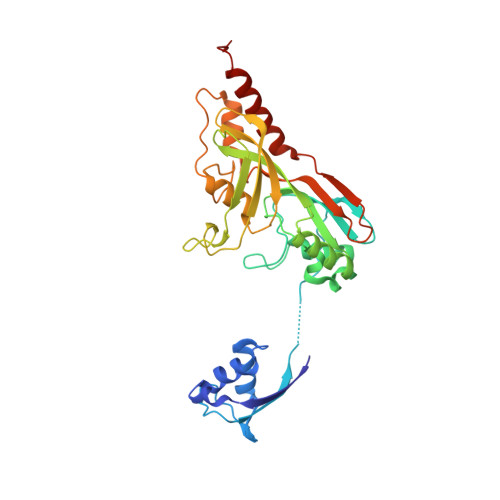 | |
UniProt | |||||
Entity Groups | |||||
| Sequence Clusters | 30% Identity50% Identity70% Identity90% Identity95% Identity100% Identity | ||||
| UniProt Group | P33643 | ||||
Sequence AnnotationsExpand | |||||
Reference Sequence | |||||
| Ligands 2 Unique | |||||
|---|---|---|---|---|---|
| ID | Chains | Name / Formula / InChI Key | 2D Diagram | 3D Interactions | |
| BCT Download:Ideal Coordinates CCD File | C [auth A], D [auth A] | BICARBONATE ION C H O3 BVKZGUZCCUSVTD-UHFFFAOYSA-M |  | ||
| CL Download:Ideal Coordinates CCD File | B [auth A] | CHLORIDE ION Cl VEXZGXHMUGYJMC-UHFFFAOYSA-M |  | ||
| Length ( Å ) | Angle ( ˚ ) |
|---|---|
| a = 67.32 | α = 90 |
| b = 75.14 | β = 90 |
| c = 85.8 | γ = 90 |
| Software Name | Purpose |
|---|---|
| REFMAC | refinement |
| PDB_EXTRACT | data extraction |
| DENZO | data reduction |
| SCALEPACK | data scaling |
| SOLVE | phasing |


