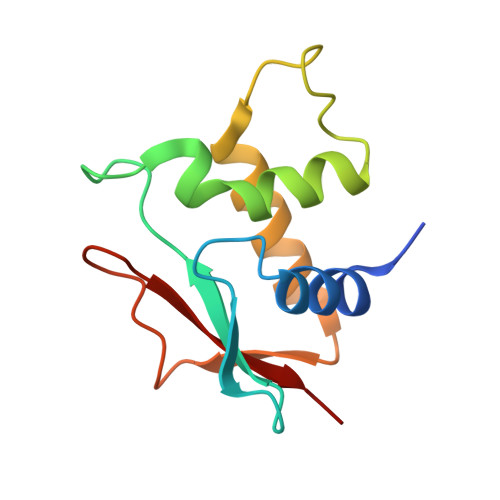
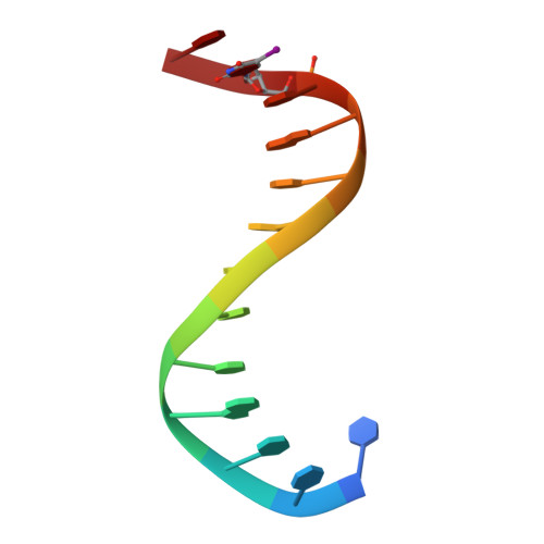
2IRF

CRYSTAL STRUCTURE OF AN IRF-2/DNA COMPLEX.


Experimental Data Snapshot

  • Method: X-RAY DIFFRACTION
  • Resolution: 2.20 Å
  • R-Value Free: 0.243 
  • R-Value Work: 0.202 
  • R-Value Observed: 0.202 

wwPDB Validation   3D Report Full Report


This is version 1.3 of the entry. See complete history


Literature

Crystal structure of an IRF-DNA complex reveals novel DNA recognition and cooperative binding to a tandem repeat of core sequences.

Fujii, Y.Shimizu, T.Kusumoto, M.Kyogoku, Y.Taniguchi, T.Hakoshima, T.

(1999) EMBO J 18: 5028-5041

  • DOI: https://doi.org/10.1093/emboj/18.18.5028
  • Primary Citation of Related Structures:  
    2IRF

  • PubMed Abstract: 

    There has been growing interest in the role of the IRF (interferon regulatory factor) family of transcription factors in the regulation of immune responses, cytokine signaling, and oncogenesis. These members are characterized by their well-conserved DNA binding domains at the N-terminal regions. Here we report the 2.2 A resolution crystal structure of the DNA binding domain of one such family member, IRF-2, bound to DNA. The structure reveals its recognition sequence, AANNGAAA (here, recognized bases are underlined and in bold, and N indicates any base), and its cooperative binding to a tandem repeat of the GAAA core sequence induced by DNA structure distortions. These facts explain well the diverse binding properties of the IRF family members, which bind to both single and tandemly repeated sequences. Furthermore, we also identified the 'helix-hairpin-strand motif' at the C terminus of the recognition helix as a metal binding site that is commonly found in certain classes of DNA-interactive proteins. Our results provide new insights into the structure and function of this family of transcription factors.


  • Organizational Affiliation

    Department of Molecular Biology, Nara Institute of Science and Technology, 8916-5 Takayama, Ikoma, Nara 630-0101, USA.


Macromolecules

Find similar proteins by:  (by identity cutoff)  |  3D Structure
Entity ID: 3
MoleculeChains Sequence LengthOrganismDetailsImage
INTERFERON REGULATORY FACTOR 2
G, H, I, J, K
G, H, I, J, K, L
113Mus musculusMutation(s): 0 
UniProt
Find proteins for P23906 (Mus musculus)
Explore P23906 
Go to UniProtKB:  P23906
Entity Groups  
Sequence Clusters30% Identity50% Identity70% Identity90% Identity95% Identity100% Identity
UniProt GroupP23906
Sequence Annotations
Expand
  • Reference Sequence

Find similar nucleic acids by:  Sequence   |   3D Structure  

Entity ID: 1
MoleculeChains LengthOrganismImage
DNA (5'-D(P*AP*AP*GP*TP*GP*AP*AP*AP*GP*(5IU)P*GP*A)-3')
A, B, C
12N/A
Sequence Annotations
Expand
  • Reference Sequence

Find similar nucleic acids by:  Sequence   |   3D Structure  

Entity ID: 2
MoleculeChains LengthOrganismImage
DNA (5'-D(*TP*TP*CP*AP*CP*TP*TP*TP*CP*AP*CP*(5IU)P*T)-3')
D, E, F
13N/A
Sequence Annotations
Expand
  • Reference Sequence
Experimental Data & Validation

Experimental Data

  • Method: X-RAY DIFFRACTION
  • Resolution: 2.20 Å
  • R-Value Free: 0.243 
  • R-Value Work: 0.202 
  • R-Value Observed: 0.202 
  • Space Group: P 65 2 2
Unit Cell:
Length ( Å )Angle ( ˚ )
a = 132.3α = 90
b = 132.3β = 90
c = 296.5γ = 120
Software Package:
Software NamePurpose
X-PLORrefinement
DENZOdata reduction
SCALEPACKdata scaling

Structure Validation

View Full Validation Report


Entry History 

Deposition Data

Revision History  (Full details and data files)

  • Version 1.0: 1999-10-08
    Type: Initial release
  • Version 1.1: 2008-04-26
    Changes: Version format compliance
  • Version 1.2: 2011-07-13
    Changes: Version format compliance
  • Version 1.3: 2023-12-27
    Changes: Data collection, Database references, Derived calculations