2IOQ | pdb_00002ioq

Crystal Structure of full-length HTPG, the Escherichia coli HSP90


Experimental Data Snapshot

  • Method: X-RAY DIFFRACTION
  • Resolution: 3.50 Å
  • R-Value Free: 
    0.370 (Depositor), 0.348 (DCC) 
  • R-Value Work: 
    0.329 (Depositor), 0.311 (DCC) 

wwPDB Validation 3D Report Full Report

Validation slider image for 2IOQ

This is version 1.3 of the entry. See complete history

Literature

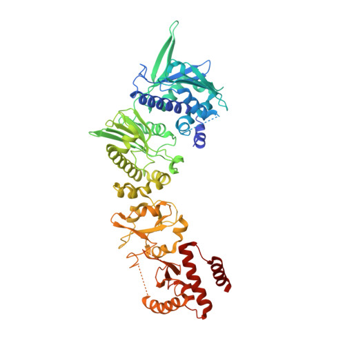
Structural Analysis of E. coli hsp90 reveals dramatic nucleotide-dependent conformational rearrangements.

Shiau, A.K.Harris, S.F.Southworth, D.R.Agard, D.A.

(2006) Cell 127: 329-340

  • DOI: https://doi.org/10.1016/j.cell.2006.09.027
  • Primary Citation Related Structures: 
    2GQ0, 2IOP, 2IOQ, 2IOR

  • PubMed Abstract: 

    In eukaryotes, the ubiquitous and abundant members of the 90 kilodalton heat-shock protein (hsp90) chaperone family facilitate the folding and conformational changes of a broad array of proteins important in cell signaling, proliferation, and survival. Here we describe the effects of nucleotides on the structure of full-length HtpG, the Escherichia coli hsp90 ortholog. By electron microscopy, the nucleotide-free, AMPPNP bound, and ADP bound states of HtpG adopt completely distinct conformations. Structural characterization of nucleotide-free and ADP bound HtpG was extended to higher resolution by X-ray crystallography. In the absence of nucleotide, HtpG exhibits an "open" conformation in which the three domains of each monomer present hydrophobic elements into the large cleft formed by the dimer. By contrast, ADP binding drives dramatic conformational changes that allow these hydrophobic elements to converge and shield each other from solvent, suggesting a mechanism by which nucleotides could control client protein binding and release.


  • Organizational Affiliation
    • Howard Hughes Medical Institute and Department of Biochemistry and Biophysics, University of California, San Francisco, 94158, USA.

Macromolecule Content 

  • Total Structure Weight: 143.04 kDa 
  • Atom Count: 9,007 
  • Modeled Residue Count: 1,144 
  • Deposited Residue Count: 1,248 
  • Unique protein chains: 1

Macromolecules

Find similar proteins by:|  3D Structure
Entity ID: 1
MoleculeChains  Sequence LengthOrganismDetailsImage
Chaperone protein htpG
A, B
624Escherichia coliMutation(s): 0 
Gene Names: htpG
UniProt
Find proteins for P0A6Z3 (Escherichia coli (strain K12))
Explore P0A6Z3 
Go to UniProtKB:  P0A6Z3
Entity Groups
Sequence Clusters30% Identity50% Identity70% Identity90% Identity95% Identity100% Identity
UniProt GroupP0A6Z3
Sequence Annotations
Expand
Reference Sequence

Experimental Data & Validation

Experimental Data

  • Method: X-RAY DIFFRACTION
  • Resolution: 3.50 Å
  • R-Value Free:  0.370 (Depositor), 0.348 (DCC) 
  • R-Value Work:  0.329 (Depositor), 0.311 (DCC) 
Space Group: P 65 2 2
Unit Cell:
Length ( Å )Angle ( ˚ )
a = 105.124α = 90
b = 105.124β = 90
c = 531.077γ = 120
Software Package:
Software NamePurpose
ADSCdata collection
CNSrefinement
DENZOdata reduction
SCALEPACKdata scaling
CNSphasing

Structure Validation

View Full Validation Report


Entry History 

Deposition Data

Revision History  (Full details and data files)

  • Version 1.0: 2006-11-21
    Type: Initial release
  • Version 1.1: 2008-05-01
    Changes: Version format compliance
  • Version 1.2: 2011-07-13
    Changes: Version format compliance
  • Version 1.3: 2024-02-21
    Changes: Data collection, Database references