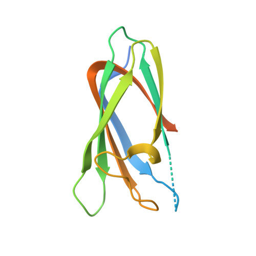
Crystal structure of the anabaena sensory rhodopsin transducer.
Vogeley, L., Trivedi, V.D., Sineshchekov, O.A., Spudich, E.N., Spudich, J.L., Luecke, H.(2007) J Mol Biology 367: 741-751
- PubMed: 17289074 Search on PubMedSearch on PubMed Central
- DOI: https://doi.org/10.1016/j.jmb.2006.11.074
- Primary Citation Related Structures:
2II7, 2II8, 2II9, 2IIA - PubMed Abstract:
We present crystal structures of the Anabaena sensory rhodopsin transducer (ASRT), a soluble cytoplasmic protein that interacts with the first structurally characterized eubacterial retinylidene photoreceptor Anabaena sensory rhodopsin (ASR). Four crystal structures of ASRT from three different spacegroups were obtained, in all of which ASRT is present as a planar (C4) tetramer, consistent with our characterization of ASRT as a tetramer in solution. The ASRT tetramer is tightly packed, with large interfaces where the well-structured beta-sandwich portion of the monomers provides the bulk of the tetramer-forming interactions, and forms a flat, stable surface on one side of the tetramer (the beta-face). Only one of our four different ASRT crystals reveals a C-terminal alpha-helix in the otherwise all-beta protein, together with a large loop from each monomer on the opposite face of the tetramer (the alpha-face), which is flexible and largely disordered in the other three crystal forms. Gel-filtration chromatography demonstrated that ASRT forms stable tetramers in solution and isothermal microcalorimetry showed that the ASRT tetramer binds to ASR with a stoichiometry of one ASRT tetramer per one ASR photoreceptor with a K(d) of 8 microM in the highest affinity measurements. Possible mechanisms for the interaction of this transducer tetramer with the ASR photoreceptor via its flexible alpha-face to mediate transduction of the light signal are discussed.
- Department of Molecular Biology and Biochemistry, University of California, Irvine, CA 92697, USA.
Organizational Affiliation: