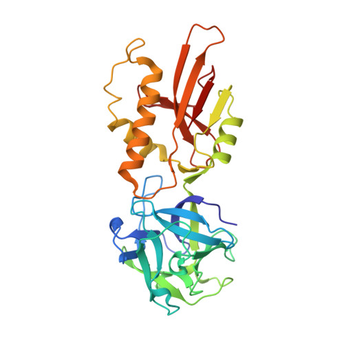
2IHO

Crystal structure of MOA, a lectin from the mushroom Marasmius oreades in complex with the trisaccharide Gal(1,3)Gal(1,4)GlcNAc


Experimental Data Snapshot

  • Method: X-RAY DIFFRACTION
  • Resolution: 2.41 Å
  • R-Value Free: 0.211 
  • R-Value Work: 0.190 
  • R-Value Observed: 0.192 

wwPDB Validation   3D Report Full Report


This is version 2.1 of the entry. See complete history


Literature

Crystal structure of the marasmius oreades mushroom lectin in complex with a xenotransplantation epitope.

Grahn, E.Askarieh, G.Holmner, A.Tateno, H.Winter, H.C.Goldstein, I.J.Krengel, U.

(2007) J Mol Biol 369: 710-721

  • DOI: https://doi.org/10.1016/j.jmb.2007.03.016
  • Primary Citation of Related Structures:  
    2IHO

  • PubMed Abstract: 

    MOA, a lectin from the mushroom Marasmius oreades, is one of the few reagents that specifically agglutinate blood group B erythrocytes. Further, it is the only lectin known to have exclusive specificity for Galalpha(1,3)Gal-containing sugar epitopes, which are antigens that pose a severe barrier to animal-to-human organ transplantation. We describe here the structure of MOA at 2.4 A resolution, in complex with the linear trisaccharide Galalpha(1,3)Galbeta(1,4)GlcNAc. The structure is dimeric, with two distinct domains per protomer: the N-terminal lectin module adopts a ricinB/beta-trefoil fold and contains three putative carbohydrate-binding sites, while the C-terminal domain serves as a dimerization interface. This latter domain, which has an unknown function, reveals a novel fold with intriguing conservation of an active site cleft. A number of indications suggest that MOA may have an enzymatic function in addition to the sugar-binding properties.


  • Organizational Affiliation

    Department of Chemistry, University of Oslo, P.O. Box 1033, Blindern, NO-0315 Oslo, Norway. elin.grahn@kjemi.uio.no


Macromolecules
Find similar proteins by:  (by identity cutoff)  |  3D Structure
Entity ID: 1
MoleculeChains Sequence LengthOrganismDetailsImage
Lectin293Marasmius oreadesMutation(s): 0 
UniProt
Find proteins for Q8X123 (Marasmius oreades)
Explore Q8X123 
Go to UniProtKB:  Q8X123
Entity Groups  
Sequence Clusters30% Identity50% Identity70% Identity90% Identity95% Identity100% Identity
UniProt GroupQ8X123
Sequence Annotations
Expand
  • Reference Sequence
Oligosaccharides

Help

Entity ID: 2
MoleculeChains Length2D Diagram Glycosylation3D Interactions
beta-D-galactopyranose-(1-3)-beta-D-galactopyranose-(1-4)-2-acetamido-2-deoxy-beta-D-glucopyranose
B, C
3N/A
Glycosylation Resources
GlyTouCan:  G35116BF
GlyCosmos:  G35116BF
GlyGen:  G35116BF
Experimental Data & Validation

Experimental Data

  • Method: X-RAY DIFFRACTION
  • Resolution: 2.41 Å
  • R-Value Free: 0.211 
  • R-Value Work: 0.190 
  • R-Value Observed: 0.192 
  • Space Group: P 32 2 1
Unit Cell:
Length ( Å )Angle ( ˚ )
a = 104.64α = 90
b = 104.64β = 90
c = 112.66γ = 120
Software Package:
Software NamePurpose
REFMACrefinement
ProDCdata collection
SHELXSphasing

Structure Validation

View Full Validation Report


Entry History 

Deposition Data

Revision History  (Full details and data files)

  • Version 1.0: 2007-05-22
    Type: Initial release
  • Version 1.1: 2008-05-01
    Changes: Version format compliance
  • Version 1.2: 2011-07-13
    Changes: Derived calculations, Version format compliance
  • Version 1.3: 2017-10-18
    Changes: Refinement description
  • Version 2.0: 2020-07-29
    Type: Remediation
    Reason: Carbohydrate remediation
    Changes: Advisory, Atomic model, Data collection, Derived calculations, Structure summary
  • Version 2.1: 2024-02-21
    Changes: Data collection, Database references, Structure summary