2IEH

Crystal structure of human kinesin Eg5 in complex with (R)-mon97, a new monastrol-based inhibitor that binds as (R)-enantiomer


Experimental Data Snapshot

  • Method: X-RAY DIFFRACTION
  • Resolution: 2.70 Å
  • R-Value Free: 0.281 
  • R-Value Work: 0.234 
  • R-Value Observed: 0.236 

wwPDB Validation   3D Report Full Report


This is version 1.3 of the entry. See complete history


Literature

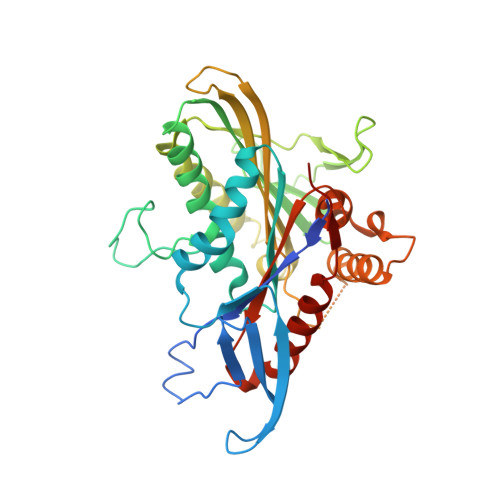
Structure of human Eg5 in complex with a new monastrol-based inhibitor bound in the R configuration.

Garcia-Saez, I.DeBonis, S.Lopez, R.Trucco, F.Rousseau, B.Thuery, P.Kozielski, F.

(2007) J Biol Chem 282: 9740-9747

  • DOI: https://doi.org/10.1074/jbc.M608883200
  • Primary Citation of Related Structures:  
    2IEH

  • PubMed Abstract: 

    Drugs that target mitotic spindle proteins have been proven useful for tackling tumor growth. Eg5, a kinesin-5 family member, represents a potential target, since its inhibition leads to prolonged mitotic arrest through the activation of the mitotic checkpoint and apoptotic cell death. Monastrol, a specific dihydropyrimidine inhibitor of Eg5, shows stereo-specificity, since predominantly the (S)-, but not the (R)-, enantiomer has been shown to be the biologically active compound in vitro and in cell-based assays. Here, we solved the crystal structure (2.7A) of the complex between human Eg5 and a new keto derivative of monastrol (named mon-97), a potent antimitotic inhibitor. Surprisingly, we identified the (R)-enantiomer bound in the active site, and not, as for monastrol, the (S)-enantiomer. The absolute configuration of this more active (R)-enantiomer has been unambiguously determined via chemical correlation and x-ray analysis. Unexpectedly, both the R- and the S-forms inhibit Eg5 ATPase activity with IC(50) values of 110 and 520 nM (basal assays) and 150 nm and 650 nm (microtubule-stimulated assays), respectively. However, the difference was large enough for the protein to select the (R)- over the (S)-enantiomer. Taken together, these results show that in this new monastrol family, both (R)- and (S)-enantiomers can be active as Eg5 inhibitors. This considerably broadens the alternatives for rational drug design.


  • Organizational Affiliation

    Laboratoire des Moteurs Moléculaires, IBS, Institut de Biologie Structurale Jean-Pierre Ebel, CNRS-Commissariat à l'Energie Atomique (CEA)-Université Joseph Fourier, 41 rue Jules Horowitz, F-38027 Grenoble.


Macromolecules
Find similar proteins by:  (by identity cutoff)  |  3D Structure
Entity ID: 1
MoleculeChains Sequence LengthOrganismDetailsImage
Kinesin-like protein KIF11
A, B
367Homo sapiensMutation(s): 0 
Gene Names: KIF11EG5KNSL1TRIP5
EC: 3.6.4.4
UniProt & NIH Common Fund Data Resources
Find proteins for P52732 (Homo sapiens)
Explore P52732 
Go to UniProtKB:  P52732
PHAROS:  P52732
Entity Groups  
Sequence Clusters30% Identity50% Identity70% Identity90% Identity95% Identity100% Identity
UniProt GroupP52732
Sequence Annotations
Expand
  • Reference Sequence
Small Molecules
Ligands 6 Unique
IDChains Name / Formula / InChI Key2D Diagram3D Interactions
ADP
Query on ADP

Download Ideal Coordinates CCD File 
F [auth A],
J [auth B]
ADENOSINE-5'-DIPHOSPHATE
C10 H15 N5 O10 P2
XTWYTFMLZFPYCI-KQYNXXCUSA-N
MOY
Query on MOY

Download Ideal Coordinates CCD File 
G [auth A],
K [auth B]
[(4R)-4-(3-HYDROXYPHENYL)-1,6-DIMETHYL-2-THIOXO-1,2,3,4-TETRAHYDROPYRIMIDIN-5-YL](PHENYL)METHANONE
C19 H18 N2 O2 S
JGBBILLMZPWNFU-QGZVFWFLSA-N
PG4
Query on PG4

Download Ideal Coordinates CCD File 
H [auth A]TETRAETHYLENE GLYCOL
C8 H18 O5
UWHCKJMYHZGTIT-UHFFFAOYSA-N
K
Query on K

Download Ideal Coordinates CCD File 
D [auth A]POTASSIUM ION
K
NPYPAHLBTDXSSS-UHFFFAOYSA-N
CL
Query on CL

Download Ideal Coordinates CCD File 
E [auth A]CHLORIDE ION
Cl
VEXZGXHMUGYJMC-UHFFFAOYSA-M
MG
Query on MG

Download Ideal Coordinates CCD File 
C [auth A],
I [auth B]
MAGNESIUM ION
Mg
JLVVSXFLKOJNIY-UHFFFAOYSA-N
Binding Affinity Annotations 
IDSourceBinding Affinity
MOY BindingDB:  2IEH IC50: min: 110, max: 150 (nM) from 2 assay(s)
PDBBind:  2IEH IC50: 110 (nM) from 1 assay(s)
Experimental Data & Validation

Experimental Data

  • Method: X-RAY DIFFRACTION
  • Resolution: 2.70 Å
  • R-Value Free: 0.281 
  • R-Value Work: 0.234 
  • R-Value Observed: 0.236 
  • Space Group: P 21 21 21
Unit Cell:
Length ( Å )Angle ( ˚ )
a = 69.37α = 90
b = 79.9β = 90
c = 159.86γ = 90
Software Package:
Software NamePurpose
Xnemodata collection
CNSrefinement
XDSdata reduction
XSCALEdata scaling
CNSphasing

Structure Validation

View Full Validation Report


Entry History 

Deposition Data

Revision History  (Full details and data files)

  • Version 1.0: 2007-01-23
    Type: Initial release
  • Version 1.1: 2008-05-01
    Changes: Version format compliance
  • Version 1.2: 2011-07-13
    Changes: Version format compliance
  • Version 1.3: 2023-08-30
    Changes: Data collection, Database references, Derived calculations, Refinement description