2ID9

1.85 A Structure of T87I/Y106W Phosphono-CheY


Experimental Data Snapshot

  • Method: X-RAY DIFFRACTION
  • Resolution: 1.75 Å
  • R-Value Free: 0.202 
  • R-Value Work: 0.170 

wwPDB Validation   3D Report Full Report


This is version 2.1 of the entry. See complete history


Literature

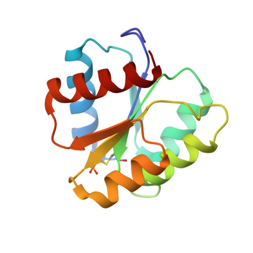
The structures of T87I phosphono-CheY and T87I/Y106W phosphono-CheY help to explain their binding affinities to the FliM and CheZ peptides.

McAdams, K.Casper, E.S.Matthew Haas, R.Santarsiero, B.D.Eggler, A.L.Mesecar, A.Halkides, C.J.

(2008) Arch Biochem Biophys 479: 105-113

  • DOI: https://doi.org/10.1016/j.abb.2008.08.019
  • Primary Citation of Related Structures:  
    2ID7, 2ID9, 2IDM

  • PubMed Abstract: 

    CheY is a response regulator in bacterial chemotaxis. Escherichia coli CheY mutants T87I and T87I/Y106W CheY are phosphorylatable on Asp57 but unable to generate clockwise rotation of the flagella. To understand this phenotype in terms of structure, stable analogs of the two CheY-P mutants were synthesized: T87I phosphono-CheY and T87I phosphono-CheY. Dissociation constants for peptides derived from flagellar motor protein FliM and phosphatase CheZ were determined for phosphono-CheY and the two mutants. The peptides bind phosphono-CheY almost as strongly as CheY-P; however, they do not bind T87I phosphono-CheY or T87I/Y106W phosphono-CheY, implying that the mutant proteins cannot bind FliM or CheZ tightly in vivo. The structures of T87I phosphono-CheY and T87I/Y106W phosphono-CheY were solved to resolutions of 1.8 and 2.4A, respectively. The increased bulk of I87 forces the side-chain of Y106 or W106, into a more solvent-accessible conformation, which occludes the peptide-binding site.


  • Organizational Affiliation

    Department of Chemistry and Biochemistry, University of North Carolina Wilmington, 601 S. College Road, Wilmington, NC 28403, USA.


Macromolecules
Find similar proteins by:  (by identity cutoff)  |  3D Structure
Entity ID: 1
MoleculeChains Sequence LengthOrganismDetailsImage
Chemotaxis protein cheY128Escherichia coliMutation(s): 3 
Gene Names: cheY
UniProt
Find proteins for P0AE67 (Escherichia coli (strain K12))
Explore P0AE67 
Go to UniProtKB:  P0AE67
Entity Groups  
Sequence Clusters30% Identity50% Identity70% Identity90% Identity95% Identity100% Identity
UniProt GroupP0AE67
Sequence Annotations
Expand
  • Reference Sequence
Small Molecules
Modified Residues  1 Unique
IDChains TypeFormula2D DiagramParent
CYQ
Query on CYQ
A
L-PEPTIDE LINKINGC4 H10 N O5 P SCYS
Experimental Data & Validation

Experimental Data

  • Method: X-RAY DIFFRACTION
  • Resolution: 1.75 Å
  • R-Value Free: 0.202 
  • R-Value Work: 0.170 
  • Space Group: P 21 21 21
Unit Cell:
Length ( Å )Angle ( ˚ )
a = 41.35α = 90
b = 49.96β = 90
c = 53.9γ = 90
Software Package:
Software NamePurpose
DENZOdata reduction
SCALEPACKdata scaling
CNSrefinement
PDB_EXTRACTdata extraction
ADSCdata collection
EPMRphasing

Structure Validation

View Full Validation Report


Entry History 

Deposition Data

Revision History  (Full details and data files)

  • Version 1.0: 2007-09-25
    Type: Initial release
  • Version 1.1: 2011-07-13
    Changes: Version format compliance
  • Version 1.2: 2014-05-28
    Changes: Other
  • Version 1.3: 2017-10-18
    Changes: Refinement description
  • Version 1.4: 2021-10-20
    Changes: Database references, Derived calculations
  • Version 2.0: 2023-07-26
    Type: Remediation
    Changes: Advisory, Atomic model, Data collection
  • Version 2.1: 2023-08-30
    Changes: Data collection, Refinement description