2ICE

CRIg bound to C3c


Experimental Data Snapshot

  • Method: X-RAY DIFFRACTION
  • Resolution: 3.10 Å
  • R-Value Free: 0.295 
  • R-Value Work: 0.236 
  • R-Value Observed: 0.239 

wwPDB Validation   3D Report Full Report


This is version 2.1 of the entry. See complete history


Literature

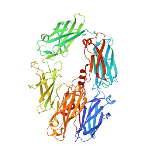
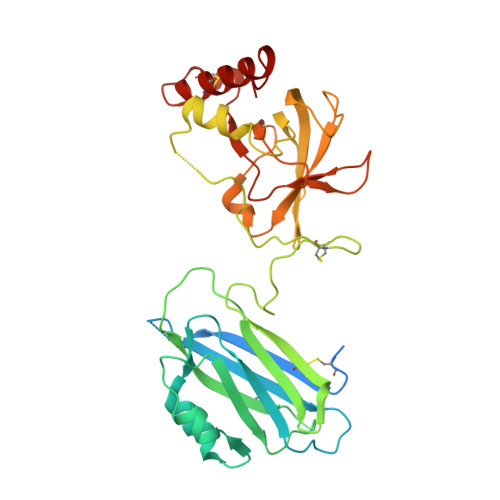
Structure of C3b in complex with CRIg gives insights into regulation of complement activation.

Wiesmann, C.Katschke, K.J.Yin, J.Helmy, K.Y.Steffek, M.Fairbrother, W.J.McCallum, S.A.Embuscado, L.DeForge, L.Hass, P.E.van Lookeren Campagne, M.

(2006) Nature 444: 217-220

  • DOI: https://doi.org/10.1038/nature05263
  • Primary Citation of Related Structures:  
    2ICC, 2ICE, 2ICF

  • PubMed Abstract: 

    The complement system is a key part of the innate immune system, and is required for clearance of pathogens from the bloodstream. After exposure to pathogens, the third component of the complement system, C3, is cleaved to C3b which, after recruitment of factor B, initiates formation of the alternative pathway convertases. CRIg, a complement receptor expressed on macrophages, binds to C3b and iC3b mediating phagocytosis of the particles, but it is unknown how CRIg selectively recognizes proteolytic C3-fragments and whether binding of CRIg to C3b inhibits convertase activation. Here we present the crystal structure of C3b in complex with CRIg and, using CRIg mutants, provide evidence that CRIg acts as an inhibitor of the alternative pathway of complement. The structure shows that activation of C3 induces major structural rearrangements, including a dramatic movement (>80 A) of the thioester-bond-containing domain through which C3b attaches to pathogen surfaces. We show that CRIg is not only a phagocytic receptor, but also a potent inhibitor of the alternative pathway convertases. The structure provides insights into the complex macromolecular structural rearrangements that occur during complement activation and inhibition. Moreover, our structure-function studies relating the structural basis of complement activation and the means by which CRIg inhibits the convertases provide important clues to the development of therapeutics that target complement.


  • Organizational Affiliation

    Department of Protein Engineering, 1 DNA Way, South San Francisco, California 94080, USA.


Macromolecules
Find similar proteins by:  (by identity cutoff)  |  3D Structure
Entity ID: 1
MoleculeChains Sequence LengthOrganismDetailsImage
Complement C3 beta chainA,
E [auth D]
642Homo sapiensMutation(s): 0 
UniProt & NIH Common Fund Data Resources
Find proteins for P01024 (Homo sapiens)
Explore P01024 
Go to UniProtKB:  P01024
PHAROS:  P01024
GTEx:  ENSG00000125730 
Entity Groups  
Sequence Clusters30% Identity50% Identity70% Identity90% Identity95% Identity100% Identity
UniProt GroupP01024
Sequence Annotations
Expand
  • Reference Sequence
Find similar proteins by:  (by identity cutoff)  |  3D Structure
Entity ID: 2
MoleculeChains Sequence LengthOrganismDetailsImage
Complement C3 alpha chainB,
F [auth E]
206Homo sapiensMutation(s): 0 
UniProt & NIH Common Fund Data Resources
Find proteins for P01024 (Homo sapiens)
Explore P01024 
Go to UniProtKB:  P01024
PHAROS:  P01024
GTEx:  ENSG00000125730 
Entity Groups  
Sequence Clusters30% Identity50% Identity70% Identity90% Identity95% Identity100% Identity
UniProt GroupP01024
Sequence Annotations
Expand
  • Reference Sequence
Find similar proteins by:  (by identity cutoff)  |  3D Structure
Entity ID: 3
MoleculeChains Sequence LengthOrganismDetailsImage
Complement C3 alpha chainC,
G [auth F]
343Homo sapiensMutation(s): 0 
UniProt & NIH Common Fund Data Resources
Find proteins for P01024 (Homo sapiens)
Explore P01024 
Go to UniProtKB:  P01024
PHAROS:  P01024
GTEx:  ENSG00000125730 
Entity Groups  
Sequence Clusters30% Identity50% Identity70% Identity90% Identity95% Identity100% Identity
UniProt GroupP01024
Sequence Annotations
Expand
  • Reference Sequence
Find similar proteins by:  (by identity cutoff)  |  3D Structure
Entity ID: 4
MoleculeChains Sequence LengthOrganismDetailsImage
V-set and immunoglobulin domain-containing protein 4D [auth S],
H [auth T]
119Homo sapiensMutation(s): 0 
Gene Names: VSIG4
UniProt & NIH Common Fund Data Resources
Find proteins for Q9Y279 (Homo sapiens)
Explore Q9Y279 
Go to UniProtKB:  Q9Y279
PHAROS:  Q9Y279
GTEx:  ENSG00000155659 
Entity Groups  
Sequence Clusters30% Identity50% Identity70% Identity90% Identity95% Identity100% Identity
UniProt GroupQ9Y279
Sequence Annotations
Expand
  • Reference Sequence
Oligosaccharides

Help

Entity ID: 5
MoleculeChains Length2D Diagram Glycosylation3D Interactions
2-acetamido-2-deoxy-beta-D-glucopyranose-(1-4)-2-acetamido-2-deoxy-beta-D-glucopyranoseI [auth G]2N-Glycosylation
Glycosylation Resources
GlyTouCan:  G42666HT
GlyCosmos:  G42666HT
GlyGen:  G42666HT
Experimental Data & Validation

Experimental Data

  • Method: X-RAY DIFFRACTION
  • Resolution: 3.10 Å
  • R-Value Free: 0.295 
  • R-Value Work: 0.236 
  • R-Value Observed: 0.239 
  • Space Group: C 1 2 1
Unit Cell:
Length ( Å )Angle ( ˚ )
a = 384.936α = 90
b = 65.213β = 102.91
c = 147.676γ = 90
Software Package:
Software NamePurpose
REFMACrefinement
PDB_EXTRACTdata extraction
HKL-2000data collection
HKL-2000data reduction
SCALEPACKdata scaling
AMoREphasing

Structure Validation

View Full Validation Report


Entry History 

Deposition Data

  • Released Date: 2006-11-07 
  • Deposition Author(s): Wiesmann, C.

Revision History  (Full details and data files)

  • Version 1.0: 2006-11-07
    Type: Initial release
  • Version 1.1: 2008-02-06
    Changes: Version format compliance
  • Version 1.2: 2011-07-13
    Changes: Advisory, Version format compliance
  • Version 1.3: 2012-09-05
    Changes: Derived calculations
  • Version 2.0: 2020-07-29
    Type: Remediation
    Reason: Carbohydrate remediation
    Changes: Atomic model, Data collection, Derived calculations, Structure summary
  • Version 2.1: 2023-08-30
    Changes: Data collection, Database references, Refinement description, Structure summary