


Structural and functional study of novel amino acid binding protein from Shigella flexneri
Fan, C.P., Wang, D.C.To be published.
Experimental Data Snapshot
wwPDB Validation 3D Report Full Report

Entity ID: 1 | |||||
|---|---|---|---|---|---|
| Molecule | Chains | Sequence Length | Organism | Details | Image |
| Putative periplasmic binding transport protein | 287 | Shigella flexneri 2a str. 301 | Mutation(s): 0 | 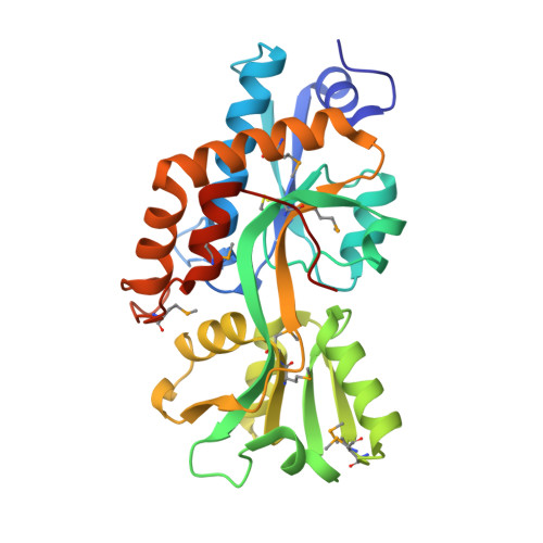 | |
UniProt | |||||
Entity Groups | |||||
| Sequence Clusters | 30% Identity50% Identity70% Identity90% Identity95% Identity100% Identity | ||||
| UniProt Group | A0A0H2UXX1 | ||||
Sequence AnnotationsExpand | |||||
Reference Sequence | |||||
| Ligands 2 Unique | |||||
|---|---|---|---|---|---|
| ID | Chains | Name / Formula / InChI Key | 2D Diagram | 3D Interactions | |
| GLU Download:Ideal Coordinates CCD File | D [auth A], F [auth B] | GLUTAMIC ACID C5 H9 N O4 WHUUTDBJXJRKMK-VKHMYHEASA-N |  | ||
| TRS Download:Ideal Coordinates CCD File | C [auth A], E [auth B] | 2-AMINO-2-HYDROXYMETHYL-PROPANE-1,3-DIOL C4 H12 N O3 LENZDBCJOHFCAS-UHFFFAOYSA-O |  | ||
| Modified Residues 1 Unique | |||||
|---|---|---|---|---|---|
| ID | Chains | Type | Formula | 2D Diagram | Parent |
| MSE Query on MSE | A, B | L-PEPTIDE LINKING | C5 H11 N O2 Se |  | MET |
| Length ( Å ) | Angle ( ˚ ) |
|---|---|
| a = 48.35 | α = 90 |
| b = 67.71 | β = 98.9 |
| c = 79.98 | γ = 90 |
| Software Name | Purpose |
|---|---|
| CNS | refinement |
| MAR345 | data collection |
| HKL-2000 | data reduction |
| HKL-2000 | data scaling |
| SOLVE | phasing |


