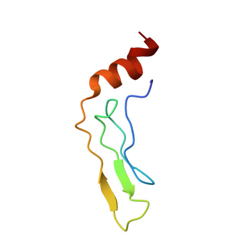
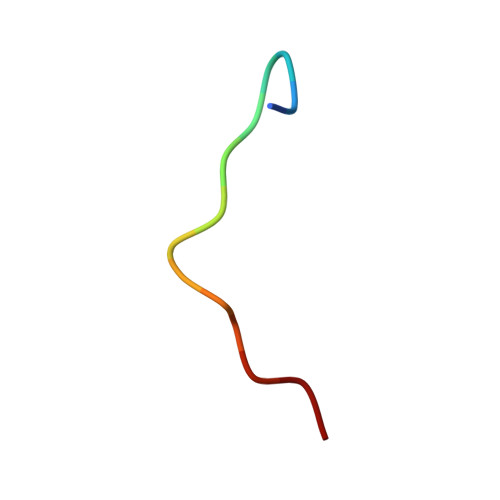
Assembly of chloroplast signal recognition particle involves structural rearrangement in cpSRP43.
Kathir, K.M., Rajalingam, D., Sivaraja, V., Kight, A., Goforth, R.L., Yu, C., Henry, R., Kumar, T.K.(2008) J Mol Biol 381: 49-60
- PubMed: 18586266 Search on PubMedSearch on PubMed Central
- DOI: https://doi.org/10.1016/j.jmb.2008.05.065
- Primary Citation Related Structures:
2HUG - PubMed Abstract:
Signal recognition particle in chloroplasts (cpSRP) exhibits the unusual ability to bind and target full-length proteins to the thylakoid membrane. Unlike cytosolic SRPs in prokaryotes and eukaryotes, cpSRP lacks an RNA moiety and functions as a heterodimer composed of a conserved 54-kDa guanosine triphosphatase (cpSRP54) and a unique 43-kDa subunit (cpSRP43). Assembly of the cpSRP heterodimer is a prerequisite for post-translational targeting activities and takes place through interactions between chromatin modifier domain 2 (CD2) of cpSRP43 and a unique 10-amino-acid region in cpSRP54 (cpSRP54(pep)). We have used multidimensional NMR spectroscopy and other biophysical methods to examine the assembly and structure of the cpSRP43-cpSRP54 interface. Our data show that CD2 of cpSRP43 binds to cpSRP54(pep) in a 1:1 stoichiometry with an apparent K(d) of approximately 1.06 muM. Steady-state fluorescence and far-UV circular dichroism data suggest that the CD2-cpSRP54(pep) interaction causes significant conformational changes in both CD2 and the peptide. Comparison of the three-dimensional solution structures of CD2 alone and in complex with cpSRP54(pep) shows that significant structural changes are induced in CD2 in order to establish a binding interface contributed mostly by residues in the N-terminal segment of CD2 (Phe5-Val10) and an arginine doublet (Arg536 and Arg537) in the cpSRP54 peptide. Taken together, our results provide new insights into the mechanism of cpSRP assembly and the structural forces that stabilize the functionally critical cpSRP43-cpSRP54 interaction.
- Department of Chemistry and Biochemistry, University of Arkansas, Fayetteville, AR 72701, USA.
Organizational Affiliation: