Monocyclic thiophenes as protein tyrosine phosphatase 1B inhibitors: Capturing interactions with Asp48.
Wan, Z.K., Lee, J., Xu, W., Erbe, D.V., Joseph-McCarthy, D., Follows, B.C., Zhang, Y.L.(2006) Bioorg Med Chem Lett 16: 4941-4945
- PubMed: 16806920 Search on PubMed
- DOI: https://doi.org/10.1016/j.bmcl.2006.06.051
- Primary Citation Related Structures:
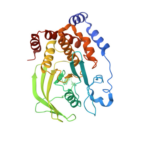
2H4G, 2H4K, 2HB1 - PubMed Abstract:
A series of monocyclic thiophenes was designed and synthesized as PTP1B inhibitors. Guided by X-ray co-crystal structural information and computational modeling, rational design led to key interactions with Asp48 and improved inhibitory potency against PTP1B.
- Chemical and Screening Sciences, Wyeth Research, 200 Cambridge Park Drive, Cambridge, MA 02140, USA.
Organizational Affiliation: