2H8K

Human Sulfotranferase SULT1C3 in complex with PAP


Experimental Data Snapshot

  • Method: X-RAY DIFFRACTION
  • Resolution: 3.20 Å
  • R-Value Free: 0.342 
  • R-Value Work: 0.292 

wwPDB Validation   3D Report Full Report


Ligand Structure Quality Assessment 


This is version 1.4 of the entry. See complete history


Literature

Structural and chemical profiling of the human cytosolic sulfotransferases.

Allali-Hassani, A.Pan, P.W.Dombrovski, L.Najmanovich, R.Tempel, W.Dong, A.Loppnau, P.Martin, F.Thornton, J.Thonton, J.Edwards, A.M.Bochkarev, A.Plotnikov, A.N.Vedadi, M.Arrowsmith, C.H.

(2007) PLoS Biol 5: e97-e97

  • DOI: https://doi.org/10.1371/journal.pbio.0050097
  • Primary Citation of Related Structures:  
    1ZD1, 2AD1, 2GWH, 2H8K

  • PubMed Abstract: 

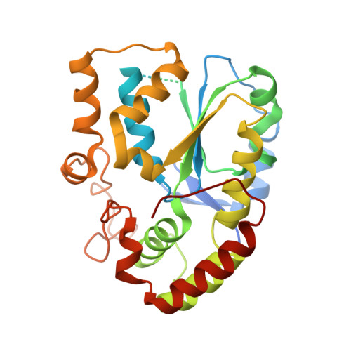
    The human cytosolic sulfotransfases (hSULTs) comprise a family of 12 phase II enzymes involved in the metabolism of drugs and hormones, the bioactivation of carcinogens, and the detoxification of xenobiotics. Knowledge of the structural and mechanistic basis of substrate specificity and activity is crucial for understanding steroid and hormone metabolism, drug sensitivity, pharmacogenomics, and response to environmental toxins. We have determined the crystal structures of five hSULTs for which structural information was lacking, and screened nine of the 12 hSULTs for binding and activity toward a panel of potential substrates and inhibitors, revealing unique "chemical fingerprints" for each protein. The family-wide analysis of the screening and structural data provides a comprehensive, high-level view of the determinants of substrate binding, the mechanisms of inhibition by substrates and environmental toxins, and the functions of the orphan family members SULT1C3 and SULT4A1. Evidence is provided for structural "priming" of the enzyme active site by cofactor binding, which influences the spectrum of small molecules that can bind to each enzyme. The data help explain substrate promiscuity in this family and, at the same time, reveal new similarities between hSULT family members that were previously unrecognized by sequence or structure comparison alone.


  • Organizational Affiliation

    Structural Genomics Consortium, University of Toronto, Toronto, Ontario, Canada.


Macromolecules
Find similar proteins by:  (by identity cutoff)  |  3D Structure
Entity ID: 1
MoleculeChains Sequence LengthOrganismDetailsImage
SULT1C3 splice variant d
A, B
306Homo sapiensMutation(s): 0 
Gene Names: SULT1C3
EC: 2.8.2
UniProt & NIH Common Fund Data Resources
Find proteins for Q6IMI6 (Homo sapiens)
Explore Q6IMI6 
Go to UniProtKB:  Q6IMI6
PHAROS:  Q6IMI6
GTEx:  ENSG00000196228 
Entity Groups  
Sequence Clusters30% Identity50% Identity70% Identity90% Identity95% Identity100% Identity
UniProt GroupQ6IMI6
Sequence Annotations
Expand
  • Reference Sequence
Small Molecules
Ligands 1 Unique
IDChains Name / Formula / InChI Key2D Diagram3D Interactions
A3P
Query on A3P

Download Ideal Coordinates CCD File 
C [auth A],
D [auth B]
ADENOSINE-3'-5'-DIPHOSPHATE
C10 H15 N5 O10 P2
WHTCPDAXWFLDIH-KQYNXXCUSA-N
Experimental Data & Validation

Experimental Data

  • Method: X-RAY DIFFRACTION
  • Resolution: 3.20 Å
  • R-Value Free: 0.342 
  • R-Value Work: 0.292 
  • Space Group: C 2 2 21
Unit Cell:
Length ( Å )Angle ( ˚ )
a = 87.161α = 90
b = 108.717β = 90
c = 136.925γ = 90
Software Package:
Software NamePurpose
DENZOdata reduction
SCALEPACKdata scaling
PHASERphasing
REFMACrefinement
PDB_EXTRACTdata extraction

Structure Validation

View Full Validation Report


Ligand Structure Quality Assessment 


Entry History 

Revision History  (Full details and data files)

  • Version 1.0: 2006-06-20
    Type: Initial release
  • Version 1.1: 2008-05-01
    Changes: Version format compliance
  • Version 1.2: 2011-07-13
    Changes: Version format compliance
  • Version 1.3: 2017-10-18
    Changes: Refinement description
  • Version 1.4: 2023-08-30
    Changes: Data collection, Database references, Derived calculations, Refinement description