2H5L | pdb_00002h5l

S-Adenosylhomocysteine hydrolase containing NAD and 3-deaza-D-eritadenine


Experimental Data Snapshot

  • Method: X-RAY DIFFRACTION
  • Resolution: 2.80 Å
  • R-Value Free: 
    0.283 (Depositor), 0.260 (DCC) 
  • R-Value Work: 
    0.248 (Depositor), 0.229 (DCC) 
  • R-Value Observed: 
    0.249 (Depositor) 

wwPDB Validation 3D Report Full Report

Validation slider image for 2H5L

Ligand Structure Quality Assessment 


This is version 1.4 of the entry. See complete history

Literature

Structure and function of eritadenine and its 3-deaza analogues: Potent inhibitors of S-adenosylhomocysteine hydrolase and hypocholesterolemic agents.

Yamada, T.Komoto, J.Lou, K.Ueki, A.Hua, D.H.Sugiyama, K.Takata, Y.Ogawa, H.Takusagawa, F.

(2007) Biochem Pharmacol 73: 981-989

  • DOI: https://doi.org/10.1016/j.bcp.2006.12.014
  • Primary Citation Related Structures: 
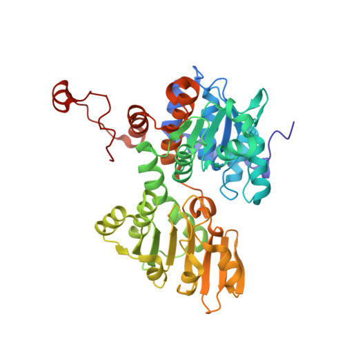
    2H5L

  • PubMed Abstract: 

    d-Eritadenine (DEA) is a potent inhibitor of S-adenosyl-l-homocysteine hydrolase (SAHH) and has hypocholesterolemic activity. We have hypothesized that 3-deaza-DEA (C3-DEA) and its analogues retain high level of SAHH inhibitory activity and have resistance to deamination and glycosidic bond hydrolysis in vivo. Such C3-DEA analogues would have much higher hypocholesterolemic activity. C3-DEA, and its methyl ester (C3-OMeDEA) and its methyl amido (C3-NMeDEA) were synthesized to examine their SAHH inhibitory and hypocholesterolemic activities. A crystal structure of SAHH containing C3-DEA was determined and confirmed that DEA and C3-DEA bound to the same site of SAHH with the same binding mode. The SAHH inhibitory activities of C3-DEA (K(I)=1.5 microM) and C3-OMeDEA (K(I)=1.5 microM) are significantly lower than that of DEA (K(I)=30 nM), while rats fed by C3-DEA and C3-OMeDEA decrease the total plasma cholesterol and phospholipids by 36-40% and 23%, respectively, which is similar to the level of reductions (42% and 27%) by DEA. C3-NMeDEA lost most of the SAHH inhibitory activity (K(I)=30 microM) and dietary C3-NMeDEA does not decrease cholesterol and phospholipid in plasma but decreases the triacylglycerol level by 16%. DEA and C3-DEA analogues are neither substrates nor inhibitors of adenosine deaminase.


  • Organizational Affiliation
    • Department of Molecular Biosciences, University of Kansas, 1200 Sunnyside Avenue, Lawrence, KS 66045, USA.

Macromolecule Content 

  • Total Structure Weight: 387.05 kDa 
  • Atom Count: 27,338 
  • Modeled Residue Count: 3,440 
  • Deposited Residue Count: 3,448 
  • Unique protein chains: 1

Macromolecules

Find similar proteins by:|  3D Structure
Entity ID: 1
MoleculeChains  Sequence LengthOrganismDetailsImage
Adenosylhomocysteinase
A, B, C, D, E
A, B, C, D, E, F, G, H
431Rattus norvegicusMutation(s): 0 
Gene Names: Ahcy
EC: 3.3.1.1 (PDB Primary Data), 3.13.2.1 (UniProt)
UniProt
Find proteins for P10760 (Rattus norvegicus)
Explore P10760 
Go to UniProtKB:  P10760
Entity Groups
Sequence Clusters30% Identity50% Identity70% Identity90% Identity95% Identity100% Identity
UniProt GroupP10760
Sequence Annotations
Expand
Reference Sequence

Small Molecules

Ligands 2 Unique
IDChains Name / Formula / InChI Key2D Diagram3D Interactions
NAD

Query on NAD


Download:Ideal Coordinates CCD File
I [auth A]
K [auth B]
M [auth C]
O [auth D]
Q [auth E]
I [auth A],
K [auth B],
M [auth C],
O [auth D],
Q [auth E],
S [auth F],
U [auth G],
W [auth H]
NICOTINAMIDE-ADENINE-DINUCLEOTIDE
C21 H27 N7 O14 P2
BAWFJGJZGIEFAR-NNYOXOHSSA-N
3DD

Query on 3DD


Download:Ideal Coordinates CCD File
J [auth A]
L [auth B]
N [auth C]
P [auth D]
R [auth E]
J [auth A],
L [auth B],
N [auth C],
P [auth D],
R [auth E],
T [auth F],
V [auth G],
X [auth H]
(2R,3R)-4-(4-AMINO-1H-IMIDAZO[4,5-C]PYRIDIN-1-YL)-2,3-DIHYDROXYBUTANOIC ACID
C10 H12 N4 O4
PBWCZGPIALILAS-HTRCEHHLSA-N

Experimental Data & Validation

Experimental Data

  • Method: X-RAY DIFFRACTION
  • Resolution: 2.80 Å
  • R-Value Free:  0.283 (Depositor), 0.260 (DCC) 
  • R-Value Work:  0.248 (Depositor), 0.229 (DCC) 
  • R-Value Observed: 0.249 (Depositor) 
Space Group: P 1 21 1
Unit Cell:
Length ( Å )Angle ( ˚ )
a = 90.5α = 90
b = 178.6β = 107.9
c = 112.66γ = 90
Software Package:
Software NamePurpose
DENZOdata reduction
X-PLORmodel building
X-PLORrefinement
SCALEPACKdata scaling
X-PLORphasing

Structure Validation

View Full Validation Report


Ligand Structure Quality Assessment 


Entry History 

Deposition Data

Revision History  (Full details and data files)

  • Version 1.0: 2007-04-10
    Type: Initial release
  • Version 1.1: 2008-05-01
    Changes: Version format compliance
  • Version 1.2: 2011-07-13
    Changes: Version format compliance
  • Version 1.3: 2017-10-18
    Changes: Refinement description
  • Version 1.4: 2024-02-14
    Changes: Data collection, Database references, Derived calculations