2H3C

Structural basis for nucleic acid and toxin recognition of the bacterial antitoxin CcdA


Experimental Data Snapshot

  • Method: SOLUTION NMR
  • Conformers Calculated: 100 
  • Conformers Submitted: 20 
  • Selection Criteria: structures with the lowest energy 

wwPDB Validation   3D Report Full Report


This is version 1.3 of the entry. See complete history


Literature

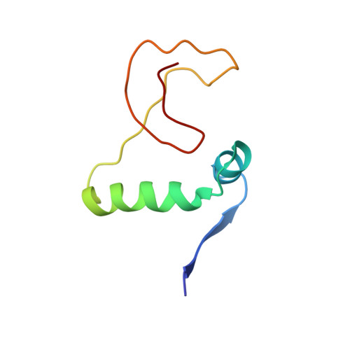
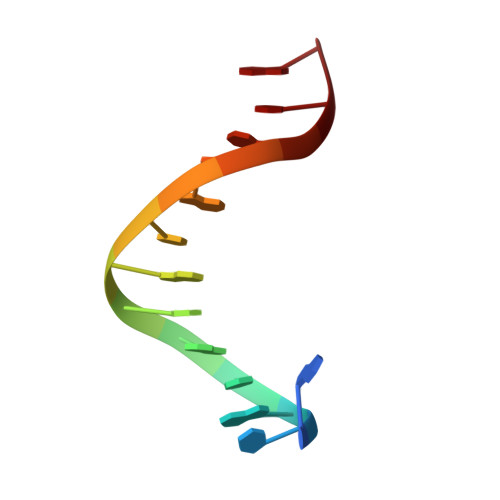
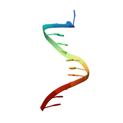
Structural Basis for Nucleic Acid and Toxin Recognition of the Bacterial Antitoxin CcdA

Madl, T.Van Melderen, L.Mine, N.Respondek, M.Oberer, M.Keller, W.Khatai, L.Zangger, K.

(2006) J Mol Biol 364: 170-185

  • DOI: https://doi.org/10.1016/j.jmb.2006.08.082
  • Primary Citation of Related Structures:  
    2ADL, 2ADN, 2H3A, 2H3C

  • PubMed Abstract: 

    Toxin-antitoxin systems are highly abundant in plasmids and bacterial chromosomes. They ensure plasmid maintenance by killing bacteria that have lost the plasmid. Their expression is autoregulated at the level of transcription. Here, we present the solution structure of CcdA, the antitoxin of the ccd system, as a free protein (16.7 kDa) and in complex with its cognate DNA (25.3 kDa). CcdA is composed of two distinct and independent domains: the N-terminal domain, responsible for DNA binding, which establishes a new family of the ribbon-helix-helix fold and the C-terminal region, which is responsible for the interaction with the toxin CcdB. The C-terminal domain is intrinsically unstructured and forms a tight complex with the toxin. We show that CcdA specifically recognizes a 6 bp palindromic DNA sequence within the operator-promoter (OP) region of the ccd operon and binds to DNA by insertion of the positively charged N-terminal beta-sheet into the major groove. The binding of up to three CcdA dimers to a 33mer DNA of its operator-promoter region was studied by NMR spectroscopy, isothermal titration calorimetry and single point mutation. The highly flexible C-terminal region of free CcdA explains its susceptibility to proteolysis by the Lon ATP-dependent protease.


  • Organizational Affiliation

    Institute of Chemistry, University of Graz, Graz 8010, Austria.


Macromolecules

Find similar proteins by:  (by identity cutoff)  |  3D Structure
Entity ID: 3
MoleculeChains Sequence LengthOrganismDetailsImage
CcdAC [auth A],
D [auth B]
72Escherichia coliMutation(s): 1 
UniProt
Find proteins for P62552 (Escherichia coli (strain K12))
Explore P62552 
Go to UniProtKB:  P62552
Entity Groups  
Sequence Clusters30% Identity50% Identity70% Identity90% Identity95% Identity100% Identity
UniProt GroupP62552
Sequence Annotations
Expand
  • Reference Sequence

Find similar nucleic acids by:  Sequence   |   3D Structure  

Entity ID: 1
MoleculeChains LengthOrganismImage
5'-D(P*AP*TP*AP*TP*GP*TP*AP*TP*AP*CP*CP*CP*G)-3'A [auth C]13N/A
Sequence Annotations
Expand
  • Reference Sequence

Find similar nucleic acids by:  Sequence   |   3D Structure  

Entity ID: 2
MoleculeChains LengthOrganismImage
5'-D(P*TP*CP*GP*GP*GP*TP*AP*TP*AP*CP*AP*TP*A)-3'B [auth D]13N/A
Sequence Annotations
Expand
  • Reference Sequence
Small Molecules
Binding Affinity Annotations 
IDSourceBinding Affinity
DNA PDBBind:  2H3C Kd: 8.85e+4 (nM) from 1 assay(s)
Experimental Data & Validation

Experimental Data

  • Method: SOLUTION NMR
  • Conformers Calculated: 100 
  • Conformers Submitted: 20 
  • Selection Criteria: structures with the lowest energy 

Structure Validation

View Full Validation Report


Entry History 

Deposition Data

Revision History  (Full details and data files)

  • Version 1.0: 2006-11-21
    Type: Initial release
  • Version 1.1: 2008-05-01
    Changes: Version format compliance
  • Version 1.2: 2011-07-13
    Changes: Version format compliance
  • Version 1.3: 2021-11-10
    Changes: Data collection, Database references, Derived calculations