2GLS | pdb_00002gls

REFINED ATOMIC MODEL OF GLUTAMINE SYNTHETASE AT 3.5 ANGSTROMS RESOLUTION


Experimental Data Snapshot

  • Method: X-RAY DIFFRACTION
  • Resolution: 3.50 Å
  • R-Value Observed: 
    0.258 (Depositor) 

wwPDB Validation 3D Report Full Report

Validation slider image for 2GLS

This is version 2.0 of the entry. See complete history

Literature

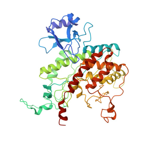
Refined atomic model of glutamine synthetase at 3.5 A resolution.

Yamashita, M.M.Almassy, R.J.Janson, C.A.Cascio, D.Eisenberg, D.

(1989) J Biological Chem 264: 17681-17690

  • DOI: https://doi.org/10.2210/pdb2gls/pdb
  • Primary Citation Related Structures: 
    2GLS

  • PubMed Abstract: 

    An atomic model of 43,692 non-hydrogen atoms has been determined for the 12-subunit enzyme glutamine synthetase from Salmonella typhimurium, by methods of x-ray diffraction including restrained least-squares atomic refinement against 65,223 unique reflections. At 3.5 A resolution the crystallographic R-factor (on 2 sigma data) is 25.8%. As reported earlier for the unrefined structure, the 12 subunits are arranged in two layers of six; at the interface of pairs of subunits within each layer, cylindrical active sites are formed by six anti-parallel beta strands contributed by one subunit and two strands by the neighboring subunit. This interpretation of the electron density map has now been supported by comparison with glutamine synthetase from Escherichia coli by the Fourier difference method. Each active site cylinder holds two Mn2+ ions, with each ion having as ligands three protein side chains and two water molecules (one water shared by both metals), as well as a histidyl side chain just beyond liganding distance. The protein ligands to Mn2+ 469 are Glu-131, Glu-212, and Glu-220; those to Mn2+ 470 are Glu-129, His-269, and Glu-357. The two layers of subunits are held together largely by the apolar COOH terminus, a helical thong, which inserts into a hydrophobic pocket formed by two neighboring subunits on the opposite ring. Also between layers, there is a hydrogen-bonded beta sheet interaction, as there is between subunits within a ring, but hydrophobic interactions account for most of the intersubunit stability. The central loop, which extends into the central aqueous channel, is subject to attack by at least five enzymes and is discussed as an enzyme "passive site."


  • Organizational Affiliation
    • Molecular Biology Institute, University of California, Los Angeles 90024.

Macromolecule Content 

  • Total Structure Weight: 623.83 kDa 
  • Atom Count: 43,692 
  • Modeled Residue Count: 5,616 
  • Deposited Residue Count: 5,628 
  • Unique protein chains: 1

Macromolecules

Find similar proteins by:|  3D Structure
Entity ID: 1
MoleculeChains  Sequence LengthOrganismDetailsImage
GLUTAMINE SYNTHETASE
A, B, C, D, E
A, B, C, D, E, F, G, H, I, J, K, L
469Salmonella enterica subsp. enterica serovar TyphimuriumMutation(s): 0 
EC: 6.3.1.2
UniProt
Find proteins for P0A1P6 (Salmonella typhimurium (strain LT2 / SGSC1412 / ATCC 700720))
Explore P0A1P6 
Go to UniProtKB:  P0A1P6
Entity Groups
Sequence Clusters30% Identity50% Identity70% Identity90% Identity95% Identity100% Identity
UniProt GroupP0A1P6
Sequence Annotations
Expand
Reference Sequence

Small Molecules

Ligands 1 Unique
IDChains Name / Formula / InChI Key2D Diagram3D Interactions
MN

Query on MN


Download:Ideal Coordinates CCD File
AA [auth H]
BA [auth H]
CA [auth I]
DA [auth I]
EA [auth J]
AA [auth H],
BA [auth H],
CA [auth I],
DA [auth I],
EA [auth J],
FA [auth J],
GA [auth K],
HA [auth K],
IA [auth L],
JA [auth L],
M [auth A],
N [auth A],
O [auth B],
P [auth B],
Q [auth C],
R [auth C],
S [auth D],
T [auth D],
U [auth E],
V [auth E],
W [auth F],
X [auth F],
Y [auth G],
Z [auth G]
MANGANESE (II) ION
Mn
WAEMQWOKJMHJLA-UHFFFAOYSA-N

Experimental Data & Validation

Experimental Data

  • Method: X-RAY DIFFRACTION
  • Resolution: 3.50 Å
  • R-Value Observed: 0.258 (Depositor) 
Space Group: C 1 2 1
Unit Cell:
Length ( Å )Angle ( ˚ )
a = 235.5α = 90
b = 134.5β = 102.8
c = 200.1γ = 90
Software Package:
Software NamePurpose
PROLSQrefinement

Structure Validation

View Full Validation Report


Entry History 

Deposition Data

Revision History  (Full details and data files)

  • Version 1.0: 1989-10-15
    Type: Initial release
  • Version 1.1: 2008-03-03
    Changes: Version format compliance
  • Version 1.2: 2011-07-13
    Changes: Advisory, Version format compliance
  • Version 2.0: 2023-09-27
    Type: Remediation
    Changes: Advisory, Atomic model, Data collection, Database references, Derived calculations, Other