


Humanization and structure of antibody clone C25
LaPorte, S.L., Finer-Moore, J., Marks, J.D., Forsyth, C.M.To be published.
Experimental Data Snapshot
wwPDB Validation 3D Report Full Report

Entity ID: 1 | |||||
|---|---|---|---|---|---|
| Molecule | Chains | Sequence Length | Organism | Details | Image |
| huC25 fab fragment light chain | 216 | Mus musculus | Mutation(s): 0 Gene Names: humanized mouse gene | 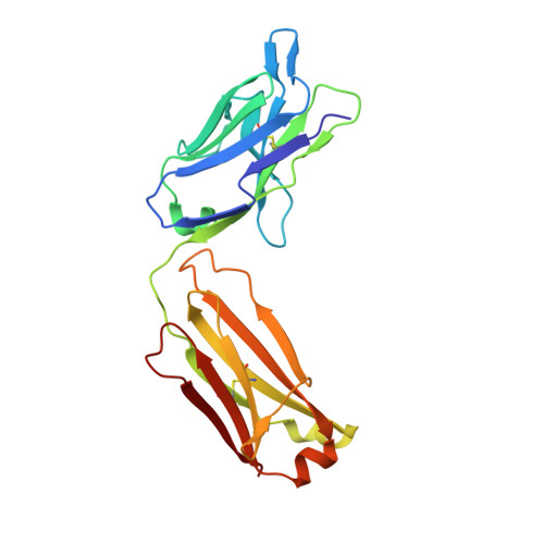 | |
Entity ID: 2 | |||||
|---|---|---|---|---|---|
| Molecule | Chains | Sequence Length | Organism | Details | Image |
| huC25 fab fragment heavy chain | 218 | Mus musculus | Mutation(s): 0 Gene Names: humanized mouse gene | 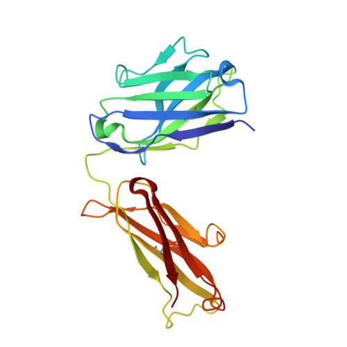 | |
| Ligands 1 Unique | |||||
|---|---|---|---|---|---|
| ID | Chains | Name / Formula / InChI Key | 2D Diagram | 3D Interactions | |
| SO4 Download:Ideal Coordinates CCD File | C [auth A], D [auth A], E [auth A], F [auth B] | SULFATE ION O4 S QAOWNCQODCNURD-UHFFFAOYSA-L |  | ||
| Length ( Å ) | Angle ( ˚ ) |
|---|---|
| a = 90.26 | α = 90 |
| b = 90.26 | β = 90 |
| c = 217.3 | γ = 120 |
| Software Name | Purpose |
|---|---|
| CNS | refinement |


