2G43

Structure of the ZNF UBP domain from deubiquitinating enzyme isopeptidase T (IsoT)


Experimental Data Snapshot

  • Method: X-RAY DIFFRACTION
  • Resolution: 2.09 Å
  • R-Value Free: 0.274 
  • R-Value Work: 0.228 
  • R-Value Observed: 0.228 

wwPDB Validation   3D Report Full Report


This is version 1.2 of the entry. See complete history


Literature

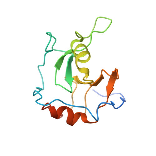
The Ubiquitin Binding Domain ZnF UBP Recognizes the C-Terminal Diglycine Motif of Unanchored Ubiquitin.

Reyes-Turcu, F.E.Horton, J.R.Mullally, J.E.Heroux, A.Cheng, X.Wilkinson, K.D.

(2006) Cell 124: 1197-1208

  • DOI: https://doi.org/10.1016/j.cell.2006.02.038
  • Primary Citation of Related Structures:  
    2G43, 2G45

  • PubMed Abstract: 

    Ubiquitin binding proteins regulate the stability, function, and/or localization of ubiquitinated proteins. Here we report the crystal structures of the zinc-finger ubiquitin binding domain (ZnF UBP) from the deubiquitinating enzyme isopeptidase T (IsoT, or USP5) alone and in complex with ubiquitin. Unlike other ubiquitin binding domains, this domain contains a deep binding pocket where the C-terminal diglycine motif of ubiquitin is inserted, thus explaining the specificity of IsoT for an unmodified C terminus on the proximal subunit of polyubiquitin. Mutations in the domain demonstrate that it is required for optimal catalytic activation of IsoT. This domain is present in several other protein families, and the ZnF UBP domain from an E3 ligase also requires the C terminus of ubiquitin for binding. These data suggest that binding the ubiquitin C terminus may be necessary for the function of other proteins.


  • Organizational Affiliation

    Department of Biochemistry, Emory University School of Medicine, Atlanta, GA 30322, USA.


Macromolecules
Find similar proteins by:  (by identity cutoff)  |  3D Structure
Entity ID: 1
MoleculeChains Sequence LengthOrganismDetailsImage
Ubiquitin carboxyl-terminal hydrolase 5
A, B
129Homo sapiensMutation(s): 0 
Gene Names: USP5ISOT
EC: 3.1.2.15
UniProt & NIH Common Fund Data Resources
Find proteins for P45974 (Homo sapiens)
Explore P45974 
Go to UniProtKB:  P45974
PHAROS:  P45974
GTEx:  ENSG00000111667 
Entity Groups  
Sequence Clusters30% Identity50% Identity70% Identity90% Identity95% Identity100% Identity
UniProt GroupP45974
Sequence Annotations
Expand
  • Reference Sequence
Experimental Data & Validation

Experimental Data

  • Method: X-RAY DIFFRACTION
  • Resolution: 2.09 Å
  • R-Value Free: 0.274 
  • R-Value Work: 0.228 
  • R-Value Observed: 0.228 
  • Space Group: C 1 2 1
Unit Cell:
Length ( Å )Angle ( ˚ )
a = 61.38α = 90
b = 86.18β = 99.29
c = 59.9γ = 90
Software Package:
Software NamePurpose
HKL-2000data collection
SCALEPACKdata scaling
SOLVEphasing
CNSrefinement
HKL-2000data reduction

Structure Validation

View Full Validation Report


Entry History 

Deposition Data

Revision History  (Full details and data files)

  • Version 1.0: 2006-04-04
    Type: Initial release
  • Version 1.1: 2008-05-01
    Changes: Version format compliance
  • Version 1.2: 2011-07-13
    Changes: Version format compliance