2G2U | pdb_00002g2u

Crystal Structure of the SHV-1 Beta-lactamase/Beta-lactamase inhibitor protein (BLIP) complex


Experimental Data Snapshot

  • Method: X-RAY DIFFRACTION
  • Resolution: 1.60 Å
  • R-Value Free: 
    0.186 (Depositor), 0.189 (DCC) 
  • R-Value Work: 
    0.173 (Depositor), 0.179 (DCC) 
  • R-Value Observed: 
    0.174 (Depositor) 

Starting Model: experimental
View more details

wwPDB Validation 3D Report Full Report

Validation slider image for 2G2U

This is version 1.5 of the entry. See complete history

Literature

Structural and Computational Characterization of the SHV-1 beta-Lactamase-beta-Lactamase Inhibitor Protein Interface.

Reynolds, K.A.Thomson, J.M.Corbett, K.D.Bethel, C.R.Berger, J.M.Kirsch, J.F.Bonomo, R.A.Handel, T.M.

(2006) J Biological Chem 281: 26745-26753

  • DOI: https://doi.org/10.1074/jbc.M603878200
  • Primary Citation Related Structures: 
    2G2U, 2G2W

  • PubMed Abstract: 

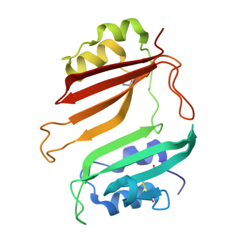
    Beta-lactamase inhibitor protein (BLIP) binds a variety of class A beta-lactamases with affinities ranging from micromolar to picomolar. Whereas the TEM-1 and SHV-1 beta-lactamases are almost structurally identical, BLIP binds TEM-1 approximately 1000-fold tighter than SHV-1. Determining the underlying source of this affinity difference is important for understanding the molecular basis of beta-lactamase inhibition and mechanisms of protein-protein interface specificity and affinity. Here we present the 1.6A resolution crystal structure of SHV-1.BLIP. In addition, a point mutation was identified, SHV D104E, that increases SHV.BLIP binding affinity from micromolar to nanomolar. Comparison of the SHV-1.BLIP structure with the published TEM-1.BLIP structure suggests that the increased volume of Glu-104 stabilizes a key binding loop in the interface. Solution of the 1.8A SHV D104K.BLIP crystal structure identifies a novel conformation in which this binding loop is removed from the interface. Using these structural data, we evaluated the ability of EGAD, a program developed for computational protein design, to calculate changes in the stability of mutant beta-lactamase.BLIP complexes. Changes in binding affinity were calculated within an error of 1.6 kcal/mol of the experimental values for 112 mutations at the TEM-1.BLIP interface and within an error of 2.2 kcal/mol for 24 mutations at the SHV-1.BLIP interface. The reasonable success of EGAD in predicting changes in interface stability is a promising step toward understanding the stability of the beta-lactamase.BLIP complexes and computationally assisted design of tight binding BLIP variants.


  • Organizational Affiliation
    • Skaggs School of Pharmacy and Pharmaceutical Sciences, University of California San Diego, La Jolla, California 92093-0684, USA.

Macromolecule Content 

  • Total Structure Weight: 46.46 kDa 
  • Atom Count: 3,590 
  • Modeled Residue Count: 430 
  • Deposited Residue Count: 430 
  • Unique protein chains: 2

Macromolecules

Find similar proteins by:|  3D Structure
Entity ID: 1
MoleculeChains  Sequence LengthOrganismDetailsImage
Beta-lactamase SHV-1265Klebsiella pneumoniaeMutation(s): 0 
Gene Names: blashv1
EC: 3.5.2.6
UniProt
Find proteins for P0AD64 (Klebsiella pneumoniae)
Explore P0AD64 
Go to UniProtKB:  P0AD64
Entity Groups
Sequence Clusters30% Identity50% Identity70% Identity90% Identity95% Identity100% Identity
UniProt GroupP0AD64
Sequence Annotations
Expand
Reference Sequence
Find similar proteins by:|  3D Structure
Entity ID: 2
MoleculeChains  Sequence LengthOrganismDetailsImage
Beta-lactamase inhibitory protein165Streptomyces clavuligerusMutation(s): 0 
UniProt
Find proteins for P35804 (Streptomyces clavuligerus)
Explore P35804 
Go to UniProtKB:  P35804
Entity Groups
Sequence Clusters30% Identity50% Identity70% Identity90% Identity95% Identity100% Identity
UniProt GroupP35804
Sequence Annotations
Expand
Reference Sequence

Experimental Data & Validation

Experimental Data

  • Method: X-RAY DIFFRACTION
  • Resolution: 1.60 Å
  • R-Value Free:  0.186 (Depositor), 0.189 (DCC) 
  • R-Value Work:  0.173 (Depositor), 0.179 (DCC) 
  • R-Value Observed: 0.174 (Depositor) 
Space Group: P 63
Unit Cell:
Length ( Å )Angle ( ˚ )
a = 127.871α = 90
b = 127.871β = 90
c = 73.4γ = 120
Software Package:
Software NamePurpose
DENZOdata reduction
SCALEPACKdata scaling
PHASERphasing
REFMACrefinement
PDB_EXTRACTdata extraction
HKL-2000data reduction

Structure Validation

View Full Validation Report


Entry History 

Deposition Data

Revision History  (Full details and data files)

  • Version 1.0: 2006-07-04
    Type: Initial release
  • Version 1.1: 2008-05-01
    Changes: Version format compliance
  • Version 1.2: 2011-07-13
    Changes: Advisory, Version format compliance
  • Version 1.3: 2017-10-18
    Changes: Refinement description
  • Version 1.4: 2023-08-30
    Changes: Data collection, Database references, Refinement description
  • Version 1.5: 2024-10-09
    Changes: Structure summary