2FUM

Catalytic domain of protein kinase PknB from Mycobacterium tuberculosis in complex with mitoxantrone


Experimental Data Snapshot

  • Method: X-RAY DIFFRACTION
  • Resolution: 2.89 Å
  • R-Value Free: 0.278 
  • R-Value Work: 0.218 
  • R-Value Observed: 0.221 

wwPDB Validation   3D Report Full Report


Ligand Structure Quality Assessment 


This is version 1.2 of the entry. See complete history


Literature

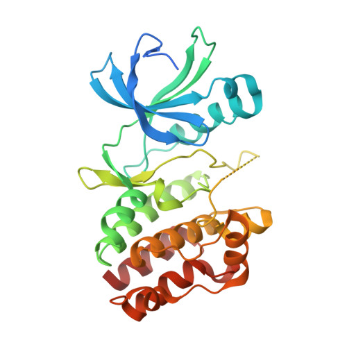
The structure of PknB in complex with mitoxantrone, an ATP-competitive inhibitor, suggests a mode of protein kinase regulation in mycobacteria

Wehenkel, A.Fernandez, P.Bellinzoni, M.Catherinot, V.Barilone, N.Labesse, G.Jackson, M.Alzari, P.M.

(2006) FEBS Lett 580: 3018-3022

  • DOI: 10.1016/j.febslet.2006.04.046

  • PubMed Abstract: 
  • Mycobacterium tuberculosis PknB is an essential receptor-like protein kinase involved in cell growth control. Here, we demonstrate that mitoxantrone, an anthraquinone derivative used in cancer therapy, is a PknB inhibitor capable of preventing mycobacterial growth ...

    Mycobacterium tuberculosis PknB is an essential receptor-like protein kinase involved in cell growth control. Here, we demonstrate that mitoxantrone, an anthraquinone derivative used in cancer therapy, is a PknB inhibitor capable of preventing mycobacterial growth. The structure of the complex reveals that mitoxantrone partially occupies the adenine-binding pocket in PknB, providing a framework for the design of compounds with potential therapeutic applications. PknB crystallizes as a 'back-to-back' homodimer identical to those observed in other structures of PknB in complex with ATP analogs. This organization resembles that of the RNA-dependent protein kinase PKR, suggesting a mechanism for kinase activation in mycobacteria.


    Organizational Affiliation

    Unité de Biochimie Structurale and CNRS URA2185, Institut Pasteur, 25 rue du Docteur Roux, 75724 Paris, France.


Macromolecules
Find similar proteins by:  (by identity cutoff)  |  3D Structure
Entity ID: 1
MoleculeChainsSequence LengthOrganismDetailsImage
Probable serine/threonine-protein kinase pknBA, B, C, D299Mycobacterium tuberculosis H37RvMutation(s): 0 
Gene Names: pknBRv0014cMTCY10H4.14c
EC: 2.7.1.37 (PDB Primary Data), 2.7.11.1 (UniProt)
UniProt
Find proteins for P9WI81 (Mycobacterium tuberculosis (strain ATCC 25618 / H37Rv))
Explore P9WI81 
Go to UniProtKB:  P9WI81
Entity Groups  
Sequence Clusters30% Identity50% Identity70% Identity90% Identity95% Identity100% Identity
UniProt GroupP9WI81
Protein Feature View
Expand
  • Reference Sequence
Small Molecules
Ligands 1 Unique
IDChainsName / Formula / InChI Key2D Diagram3D Interactions
MIX
Query on MIX

Download Ideal Coordinates CCD File 
E [auth A],
F [auth B],
G [auth C],
H [auth D]
1,4-DIHYDROXY-5,8-BIS({2-[(2-HYDROXYETHYL)AMINO]ETHYL}AMINO)-9,10-ANTHRACENEDIONE
C22 H28 N4 O6
KKZJGLLVHKMTCM-UHFFFAOYSA-N
 Ligand Interaction
Binding Affinity Annotations 
IDSourceBinding Affinity
MIX PDBBind:  2FUM IC50: 800 (nM) from 1 assay(s)
Experimental Data & Validation

Experimental Data

  • Method: X-RAY DIFFRACTION
  • Resolution: 2.89 Å
  • R-Value Free: 0.278 
  • R-Value Work: 0.218 
  • R-Value Observed: 0.221 
  • Space Group: P 43 21 2
Unit Cell:
Length ( Å )Angle ( ˚ )
a = 116.863α = 90
b = 116.863β = 90
c = 260.347γ = 90
Software Package:
Software NamePurpose
REFMACrefinement
MOSFLMdata reduction
CCP4data scaling
PHASERphasing

Structure Validation

View Full Validation Report


Ligand Structure Quality Assessment 


Entry History 

Deposition Data

Revision History  (Full details and data files)

  • Version 1.0: 2006-08-01
    Type: Initial release
  • Version 1.1: 2008-05-01
    Changes: Version format compliance
  • Version 1.2: 2011-07-13
    Changes: Advisory, Version format compliance