Refined high-resolution structure of the metal-ion dependent L-fuculose-1-phosphate aldolase (class II) from Escherichia coli.
Dreyer, M.K., Schulz, G.E.(1996) Acta Crystallogr D Biol Crystallogr 52: 1082-1091
- PubMed: 15299567 Search on PubMed
- DOI: https://doi.org/10.1107/S0907444996009146
- Primary Citation Related Structures:
1FUA, 2FUA - PubMed Abstract:
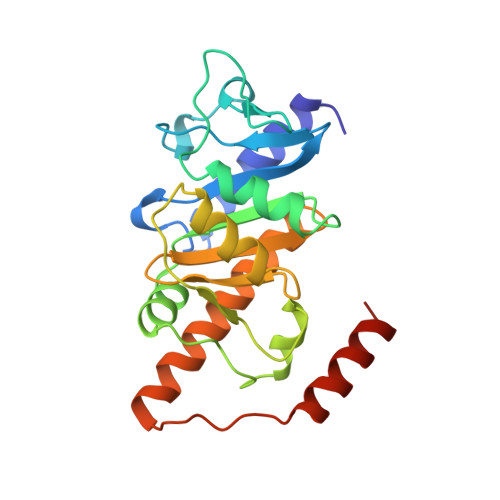
The structure of the class II zinc-ion dependent L-fuculose-1-phosphate aldolase from Escherichia coli in its tetragonal crystal form has been established at 1.92 A resolution. The homotetrameric enzyme has a molecular mass of 4 x 24 kDa and follows C(4) symmetry. The structure model is exactly symmetrical, which contradicts an observed birefringence anomaly of the crystals. The four catalytic centers are located in deep clefts at the interfaces of adjacent subunits. The zinc ion is coordinated by three histidines and one glutamate in an almost tetrahedral arrangement. In contrast to numerous other catalytically competent zinc ions, there is no water molecule in the ligand sphere. Replacement of zinc by a cobalt ion caused only small structural changes. A search through the Protein Data Bank indicated that the chain fold is novel. Sequence homology searches revealed a significant similarity to the bacterial L-ribulose-5-phosphate 4-epimerase.
- Institut für Organische Chemie und Biochemie, Albert-Ludwigs-Universität, Freiburg im Breisgau, Germany.
Organizational Affiliation: