Crystal structure of PhnH: an essential component of carbon-phosphorus lyase in Escherichia coli.
Adams, M.A., Luo, Y., Hove-Jensen, B., He, S.M., van Staalduinen, L.M., Zechel, D.L., Jia, Z.(2008) J Bacteriol 190: 1072-1083
- PubMed: 17993513 Search on PubMedSearch on PubMed Central
- DOI: https://doi.org/10.1128/JB.01274-07
- Primary Citation Related Structures:
2FSU - PubMed Abstract:
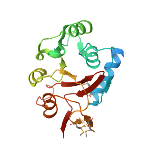
Organophosphonates are reduced forms of phosphorous that are characterized by the presence of a stable carbon-phosphorus (C-P) bond, which resists chemical hydrolysis, thermal decomposition, and photolysis. The chemically inert nature of the C-P bond has raised environmental concerns as toxic phosphonates accumulate in a number of ecosystems. Carbon-phosphorous lyase (CP lyase) is a multienzyme pathway encoded by the phn operon in gram-negative bacteria. In Escherichia coli 14 cistrons comprise the operon (phnCDEFGHIJKLMNOP) and collectively allow the internalization and degradation of phosphonates. Here we report the X-ray crystal structure of the PhnH component at 1.77 A resolution. The protein exhibits a novel fold, although local similarities with the pyridoxal 5'-phosphate-dependent transferase family of proteins are apparent. PhnH forms a dimer in solution and in the crystal structure, the interface of which is implicated in creating a potential ligand binding pocket. Our studies further suggest that PhnH may be capable of binding negatively charged cyclic compounds through interaction with strictly conserved residues. Finally, we show that PhnH is essential for C-P bond cleavage in the CP lyase pathway.
- Deparment of Biochemistry, Queen's University, Kingston, Ontario, Canada K7L3N6.
Organizational Affiliation: