2FKM

PMM/PGM S108D mutant with alpha-d-glucose 1,6-bisphosphate bound


Experimental Data Snapshot

  • Method: X-RAY DIFFRACTION
  • Resolution: 1.90 Å
  • R-Value Free: 0.251 
  • R-Value Work: 0.203 
  • R-Value Observed: 0.205 

wwPDB Validation   3D Report Full Report


Ligand Structure Quality Assessment 


This is version 1.6 of the entry. See complete history


Literature

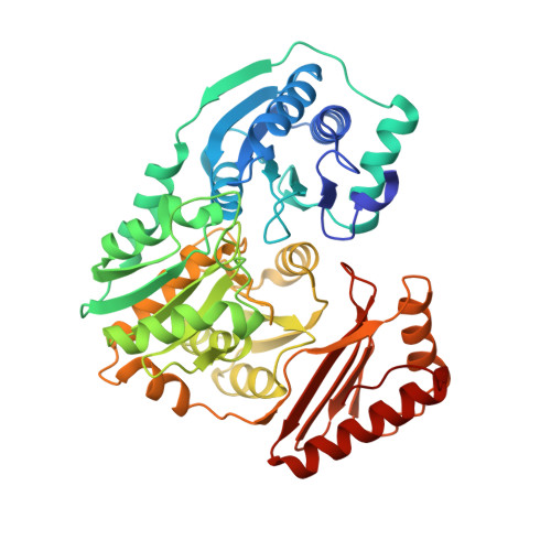
The reaction of phosphohexomutase from Pseudomonas aeruginosa: structural insights into a simple processive enzyme.

Regni, C.Schramm, A.M.Beamer, L.J.

(2006) J Biol Chem 281: 15564-15571

  • DOI: https://doi.org/10.1074/jbc.M600590200
  • Primary Citation of Related Structures:  
    2FKF, 2FKM

  • PubMed Abstract: 

    The enzyme phosphomannomutase/phosphoglucomutase (PMM/PGM) from Pseudomonas aeruginosa catalyzes the reversible conversion of 1-phospho to 6-phospho-sugars. The reaction entails two phosphoryl transfers, with an intervening 180 degrees reorientation of the reaction intermediate (e.g. glucose 1,6-bisphosphate) during catalysis. Reorientation of the intermediate occurs without dissociation from the active site of the enzyme and is, thus, a simple example of processivity, as defined by multiple rounds of catalysis without release of substrate. Structural characterization of two PMM/PGM-intermediate complexes with glucose 1,6-bisphosphate provides new insights into the reaction catalyzed by the enzyme, including the reorientation of the intermediate. Kinetic analyses of site-directed mutants prompted by the structural studies reveal active site residues critical for maintaining association with glucose 1,6-bisphosphate during its unique dynamic reorientation in the active site of PMM/PGM.


  • Organizational Affiliation

    Department of Biochemistry, University of Missouri, Columbia, Missouri 65211, USA.


Macromolecules
Find similar proteins by:  (by identity cutoff)  |  3D Structure
Entity ID: 1
MoleculeChains Sequence LengthOrganismDetailsImage
Phosphomannomutase/phosphoglucomutaseA [auth X]462Pseudomonas aeruginosaMutation(s): 1 
Gene Names: AlgC
EC: 5.4.2.8
UniProt
Find proteins for P26276 (Pseudomonas aeruginosa (strain ATCC 15692 / DSM 22644 / CIP 104116 / JCM 14847 / LMG 12228 / 1C / PRS 101 / PAO1))
Explore P26276 
Go to UniProtKB:  P26276
Entity Groups  
Sequence Clusters30% Identity50% Identity70% Identity90% Identity95% Identity100% Identity
UniProt GroupP26276
Sequence Annotations
Expand
  • Reference Sequence
Small Molecules
Ligands 2 Unique
IDChains Name / Formula / InChI Key2D Diagram3D Interactions
G16
Query on G16

Download Ideal Coordinates CCD File 
B [auth X]1,6-di-O-phosphono-alpha-D-glucopyranose
C6 H13 O12 P2
RWHOZGRAXYWRNX-VFUOTHLCSA-M
ZN
Query on ZN

Download Ideal Coordinates CCD File 
C [auth X]ZINC ION
Zn
PTFCDOFLOPIGGS-UHFFFAOYSA-N
Experimental Data & Validation

Experimental Data

  • Method: X-RAY DIFFRACTION
  • Resolution: 1.90 Å
  • R-Value Free: 0.251 
  • R-Value Work: 0.203 
  • R-Value Observed: 0.205 
  • Space Group: P 21 21 21
Unit Cell:
Length ( Å )Angle ( ˚ )
a = 70.222α = 90
b = 70.356β = 90
c = 84.392γ = 90
Software Package:
Software NamePurpose
REFMACrefinement
CNSrefinement
d*TREKdata scaling
PDB_EXTRACTdata extraction
DENZOdata reduction
CNSphasing

Structure Validation

View Full Validation Report


Ligand Structure Quality Assessment 


Entry History 

Deposition Data

Revision History  (Full details and data files)

  • Version 1.0: 2006-04-04
    Type: Initial release
  • Version 1.1: 2008-05-01
    Changes: Version format compliance
  • Version 1.2: 2011-07-13
    Changes: Advisory, Version format compliance
  • Version 1.3: 2019-07-24
    Changes: Data collection, Refinement description
  • Version 1.4: 2020-07-29
    Type: Remediation
    Reason: Carbohydrate remediation
    Changes: Data collection, Derived calculations, Structure summary
  • Version 1.5: 2021-10-20
    Changes: Database references, Structure summary
  • Version 1.6: 2023-08-30
    Changes: Data collection, Refinement description