2FF6

Crystal structure of Gelsolin domain 1:ciboulot domain 2 hybrid in complex with actin


Experimental Data Snapshot

  • Method: X-RAY DIFFRACTION
  • Resolution: 2.05 Å
  • R-Value Free: 0.237 
  • R-Value Work: 0.202 
  • R-Value Observed: 0.204 

wwPDB Validation   3D Report Full Report


Ligand Structure Quality Assessment 


This is version 1.3 of the entry. See complete history


Literature

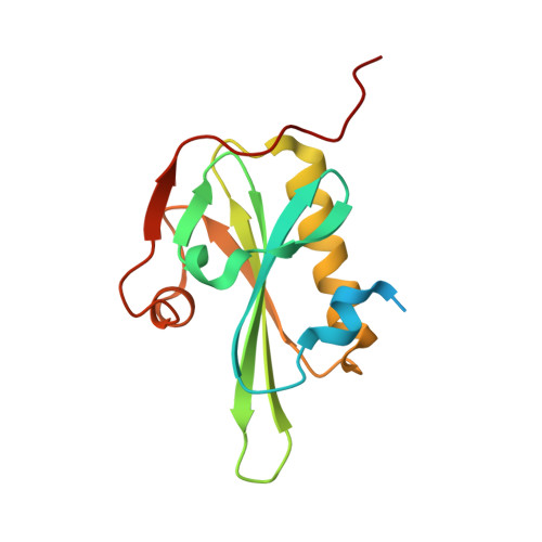
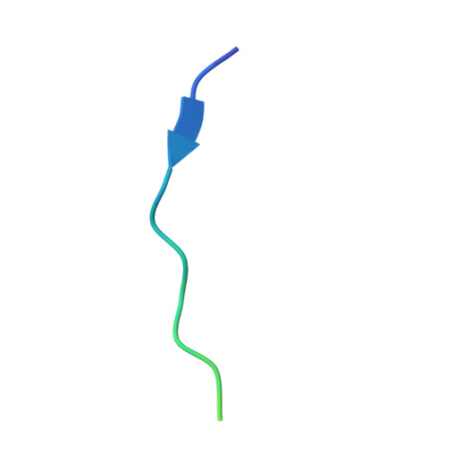
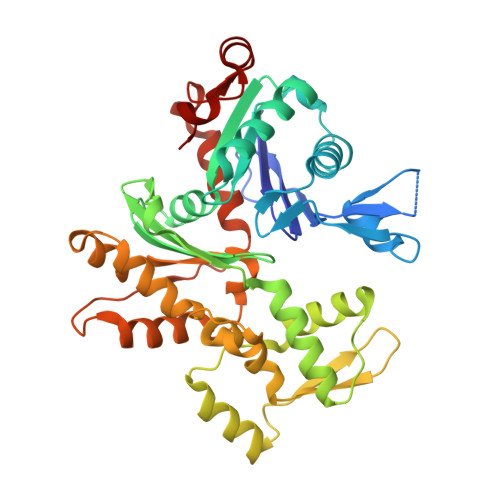
The Structural Basis of Actin Interaction with Multiple WH2/beta-Thymosin Motif-Containing Proteins

Aguda, A.H.Xue, B.Irobi, E.Preat, T.Robinson, R.C.

(2006) Structure 14: 469-476

  • DOI: https://doi.org/10.1016/j.str.2005.12.011
  • Primary Citation of Related Structures:  
    2FF3, 2FF6

  • PubMed Abstract: 
  • Participation of actin in cellular processes relies on the dynamics of filament assembly. Filament elongation is fed by monomeric actin in complex with either profilin or a Wiscott-Aldrich syndrome protein (WASP) homology domain 2 (WH2)/beta-thymosin (betaT) domain ...

    Participation of actin in cellular processes relies on the dynamics of filament assembly. Filament elongation is fed by monomeric actin in complex with either profilin or a Wiscott-Aldrich syndrome protein (WASP) homology domain 2 (WH2)/beta-thymosin (betaT) domain. WH2/betaT motif repetition (typified by ciboulot) or combination with nonrelated domains (as found in N-WASP) results in proteins that yield their actin to filament elongation. Here, we report the crystal structures of actin bound hybrid proteins, constructed between gelsolin and WH2/betaT domains from ciboulot or N-WASP. We observe the C-terminal half of ciboulot domain 2 bound to actin. In solution, we show that cibolout domains 2 and 3 bind to both G- and F-actin, and that whole ciboulot forms a complex with two actin monomers. In contrast, the analogous portion of N-WASP WH2 domain 2 is detached from actin, indicating that the C-terminal halves of the betaT and WH2 motifs are not functionally analogous.


    Organizational Affiliation

    Department of Medical Biochemistry and Microbiology, Uppsala Biomedical Center, Uppsala University, Uppsala 751 23, Sweden.


Macromolecules
Find similar proteins by:  (by identity cutoff)  |  3D Structure
Entity ID: 1
MoleculeChainsSequence LengthOrganismDetailsImage
GelsolinA [auth G]150Homo sapiensMutation(s): 0 
Gene Names: GSN
UniProt & NIH Common Fund Data Resources
Find proteins for P06396 (Homo sapiens)
Explore P06396 
Go to UniProtKB:  P06396
PHAROS:  P06396
Entity Groups  
Sequence Clusters30% Identity50% Identity70% Identity90% Identity95% Identity100% Identity
UniProt GroupP06396
Protein Feature View
Expand
  • Reference Sequence
Find similar proteins by:  (by identity cutoff)  |  3D Structure
Entity ID: 2
MoleculeChainsSequence LengthOrganismDetailsImage
CG4944-PC, isoform CB [auth H]26Drosophila melanogasterMutation(s): 0 
Gene Names: cibcib-RCDmel\CG4944EG0007-11EG:EG0007.11CG4944Dmel_CG4944
UniProt
Find proteins for Q8IRS7 (Drosophila melanogaster)
Explore Q8IRS7 
Go to UniProtKB:  Q8IRS7
Entity Groups  
Sequence Clusters30% Identity50% Identity70% Identity90% Identity95% Identity100% Identity
UniProt GroupQ8IRS7
Protein Feature View
Expand
  • Reference Sequence
Find similar proteins by:  (by identity cutoff)  |  3D Structure
Entity ID: 3
MoleculeChainsSequence LengthOrganismDetailsImage
Actin, alpha skeletal muscleC [auth A]375Oryctolagus cuniculusMutation(s): 0 
Gene Names: ACTA1ACTA
UniProt
Find proteins for P68135 (Oryctolagus cuniculus)
Explore P68135 
Go to UniProtKB:  P68135
Entity Groups  
Sequence Clusters30% Identity50% Identity70% Identity90% Identity95% Identity100% Identity
UniProt GroupP68135
Protein Feature View
Expand
  • Reference Sequence
Small Molecules
Ligands 2 Unique
IDChainsName / Formula / InChI Key2D Diagram3D Interactions
ATP
Query on ATP

Download Ideal Coordinates CCD File 
G [auth A]ADENOSINE-5'-TRIPHOSPHATE
C10 H16 N5 O13 P3
ZKHQWZAMYRWXGA-KQYNXXCUSA-N
 Ligand Interaction
CA
Query on CA

Download Ideal Coordinates CCD File 
D [auth G],
E [auth G],
F [auth A]
CALCIUM ION
Ca
BHPQYMZQTOCNFJ-UHFFFAOYSA-N
 Ligand Interaction
Experimental Data & Validation

Experimental Data

  • Method: X-RAY DIFFRACTION
  • Resolution: 2.05 Å
  • R-Value Free: 0.237 
  • R-Value Work: 0.202 
  • R-Value Observed: 0.204 
  • Space Group: P 1 21 1
Unit Cell:
Length ( Å )Angle ( ˚ )
a = 56.221α = 90
b = 69.373β = 95.1
c = 79.65γ = 90
Software Package:
Software NamePurpose
REFMACrefinement
MOSFLMdata reduction
CCP4data scaling
AMoREphasing

Structure Validation

View Full Validation Report


Ligand Structure Quality Assessment 


Entry History 

Deposition Data

Revision History  (Full details and data files)

  • Version 1.0: 2006-03-21
    Type: Initial release
  • Version 1.1: 2008-05-01
    Changes: Version format compliance
  • Version 1.2: 2011-07-13
    Changes: Version format compliance
  • Version 1.3: 2018-05-23
    Changes: Data collection