2F2K

Aldose reductase tertiary complex with NADPH and DEG


Experimental Data Snapshot

  • Method: X-RAY DIFFRACTION
  • Resolution: 1.94 Å
  • R-Value Free: 0.256 
  • R-Value Work: 0.196 
  • R-Value Observed: 0.196 

wwPDB Validation   3D Report Full Report


Ligand Structure Quality Assessment 


This is version 1.2 of the entry. See complete history


Literature

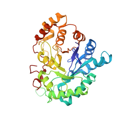
Structure of a glutathione conjugate bound to the active site of aldose reductase.

Singh, R.White, M.A.Ramana, K.V.Petrash, J.M.Watowich, S.J.Bhatnagar, A.Srivastava, S.K.

(2006) Proteins 64: 101-110

  • DOI: 10.1002/prot.20988
  • Primary Citation of Related Structures:  
    2F2K

  • PubMed Abstract: 
  • Aldose reductase (AR) is a monomeric NADPH-dependent oxidoreductase that catalyzes the reduction of aldehydes, ketones, and aldo-sugars. AR has been linked to the development of hyperglycemic injury and is a clinical target for the treatment of secondary diabetic complications ...

    Aldose reductase (AR) is a monomeric NADPH-dependent oxidoreductase that catalyzes the reduction of aldehydes, ketones, and aldo-sugars. AR has been linked to the development of hyperglycemic injury and is a clinical target for the treatment of secondary diabetic complications. In addition to reducing glucose, AR is key regulator of cell signaling through it's reduction of aldehydes derived from lipoproteins and membrane phospholipids. AR catalyzes the reduction of glutathione conjugates of unsaturated aldehydes with higher catalytic efficiency than free aldehydes. The X-ray structure of human AR holoenzyme in complex with the glutathione analogue S-(1,2-dicarboxyethyl) glutathione (DCEG) was determined at a resolution of 1.94 A. The distal carboxylate group of DCEG's dicarboxyethyl moiety interacted with the conserved AR anion binding site residues Tyr48, His110, and Trp111. The bound DCEG's glutathione backbone adopted the low-energy Y-shape form. The C-terminal carboxylate of DCEG glutathione's glycine formed hydrogen bonds to Leu301 and Ser302, while the remaining interactions between DCEG and AR were hydrophobic, permitting significant flexibility of the AR and glutathione (GS) analogue interaction. The observed conformation and interactions of DCEG with AR were consistent with our previously published molecular dynamics model of glutathionyl-propanal binding to AR. The current structure identifies major interactions of glutathione conjugates with the AR active-site residues.


    Organizational Affiliation

    Department of Biochemistry and Molecular Biology, University of Texas Medical Branch, Galveston 77555-0647, USA.


Macromolecules
Find similar proteins by:  (by identity cutoff)  |  3D Structure
Entity ID: 1
MoleculeChainsSequence LengthOrganismDetailsImage
Aldose reductaseA316Homo sapiensMutation(s): 0 
Gene Names: AKR1B1ALDR1ALR2
EC: 1.1.1.21 (PDB Primary Data), 1.1.1.300 (UniProt), 1.1.1.372 (UniProt), 1.1.1.54 (UniProt)
UniProt & NIH Common Fund Data Resources
Find proteins for P15121 (Homo sapiens)
Explore P15121 
Go to UniProtKB:  P15121
PHAROS:  P15121
Entity Groups  
Sequence Clusters30% Identity50% Identity70% Identity90% Identity95% Identity100% Identity
UniProt GroupP15121
Protein Feature View
Expand
  • Reference Sequence
Small Molecules
Ligands 2 Unique
IDChainsName / Formula / InChI Key2D Diagram3D Interactions
NDP
Query on NDP

Download Ideal Coordinates CCD File 
B [auth A]NADPH DIHYDRO-NICOTINAMIDE-ADENINE-DINUCLEOTIDE PHOSPHATE
C21 H30 N7 O17 P3
ACFIXJIJDZMPPO-NNYOXOHSSA-N
 Ligand Interaction
TGG
Query on TGG

Download Ideal Coordinates CCD File 
C [auth A]GAMMA-GLUTAMYL-S-(1,2-DICARBOXYETHYL)CYSTEINYLGLYCINE
C14 H21 N3 O10 S
PWCIUOASSAHGHI-BIIVOSGPSA-N
 Ligand Interaction
Experimental Data & Validation

Experimental Data

  • Method: X-RAY DIFFRACTION
  • Resolution: 1.94 Å
  • R-Value Free: 0.256 
  • R-Value Work: 0.196 
  • R-Value Observed: 0.196 
  • Space Group: P 1 21 1
Unit Cell:
Length ( Å )Angle ( ˚ )
a = 47.2α = 90
b = 66.7β = 92.25
c = 49.3γ = 90
Software Package:
Software NamePurpose
DENZOdata reduction
SCALEPACKdata scaling
CNSrefinement
CNSphasing

Structure Validation

View Full Validation Report


Ligand Structure Quality Assessment 


Entry History 

Deposition Data

Revision History  (Full details and data files)

  • Version 1.0: 2006-05-16
    Type: Initial release
  • Version 1.1: 2008-05-01
    Changes: Version format compliance
  • Version 1.2: 2011-07-13
    Changes: Version format compliance