2F2E

Crystal Structure of PA1607, a Putative Transcription Factor


Experimental Data Snapshot

  • Method: X-RAY DIFFRACTION
  • Resolution: 1.85 Å
  • R-Value Free: 0.251 
  • R-Value Work: 0.177 
  • R-Value Observed: 0.181 

wwPDB Validation   3D Report Full Report


Ligand Structure Quality Assessment 


This is version 1.3 of the entry. See complete history


Literature

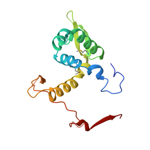
The X-ray crystal structure of PA1607 from Pseudomonas aureginosa at 1.9 A resolution--a putative transcription factor.

Sieminska, E.A.Xu, X.Savchenko, A.Sanders, D.A.

(2007) Protein Sci 16: 543-549

  • DOI: https://doi.org/10.1110/ps.062668207
  • Primary Citation of Related Structures:  
    2F2E

  • PubMed Abstract: 
  • The structure of the PA1607 protein from Pseudomonas aureginosa was determined at 1.85 A resolution using the Se-Met multiwavelength anomalous diffraction (MAD) technique. PA1607 forms a dimer and adopts a winged-helix motif similar to the MarR family of transcription regulators, though it has an unusual dimerization profile ...

    The structure of the PA1607 protein from Pseudomonas aureginosa was determined at 1.85 A resolution using the Se-Met multiwavelength anomalous diffraction (MAD) technique. PA1607 forms a dimer and adopts a winged-helix motif similar to the MarR family of transcription regulators, though it has an unusual dimerization profile. The DNA-binding regions and a putative metal-binding site are not conserved in PA1607.


    Organizational Affiliation

    Department of Chemistry, University of Saskatchewan, Saskatoon, Saskatchewan S7N 5C9, Canada.


Macromolecules
Find similar proteins by:  (by identity cutoff)  |  3D Structure
Entity ID: 1
MoleculeChainsSequence LengthOrganismDetailsImage
PA1607
A, B
146Pseudomonas aeruginosaMutation(s): 3 
Gene Names: PA1607
UniProt
Find proteins for Q9I3B4 (Pseudomonas aeruginosa (strain ATCC 15692 / DSM 22644 / CIP 104116 / JCM 14847 / LMG 12228 / 1C / PRS 101 / PAO1))
Explore Q9I3B4 
Go to UniProtKB:  Q9I3B4
Entity Groups  
Sequence Clusters30% Identity50% Identity70% Identity90% Identity95% Identity100% Identity
UniProt GroupQ9I3B4
Protein Feature View
Expand
  • Reference Sequence
Small Molecules
Ligands 2 Unique
IDChainsName / Formula / InChI Key2D Diagram3D Interactions
GLC
Query on GLC

Download Ideal Coordinates CCD File 
E [auth B]alpha-D-glucopyranose
C6 H12 O6
WQZGKKKJIJFFOK-DVKNGEFBSA-N
 Ligand Interaction
SO4
Query on SO4

Download Ideal Coordinates CCD File 
C [auth A],
D [auth A],
F [auth B],
G [auth B]
SULFATE ION
O4 S
QAOWNCQODCNURD-UHFFFAOYSA-L
 Ligand Interaction
Modified Residues  1 Unique
IDChainsTypeFormula2D DiagramParent
MSE
Query on MSE
A, B
L-PEPTIDE LINKINGC5 H11 N O2 SeMET
Experimental Data & Validation

Experimental Data

  • Method: X-RAY DIFFRACTION
  • Resolution: 1.85 Å
  • R-Value Free: 0.251 
  • R-Value Work: 0.177 
  • R-Value Observed: 0.181 
  • Space Group: C 1 2 1
Unit Cell:
Length ( Å )Angle ( ˚ )
a = 46.884α = 90
b = 78.865β = 91.64
c = 78.941γ = 90
Software Package:
Software NamePurpose
REFMACrefinement
HKL-2000data reduction
SCALEPACKdata scaling
SOLVEphasing

Structure Validation

View Full Validation Report


Ligand Structure Quality Assessment 


Entry History 

Revision History  (Full details and data files)

  • Version 1.0: 2006-03-14
    Type: Initial release
  • Version 1.1: 2008-05-01
    Changes: Version format compliance
  • Version 1.2: 2011-07-13
    Changes: Version format compliance
  • Version 1.3: 2020-07-29
    Type: Remediation
    Reason: Carbohydrate remediation
    Changes: Advisory, Data collection, Database references, Derived calculations, Structure summary